
Table of Contents
The invention of the microscope fundamentally changed how humanity understands the world. By granting us the ability to look beyond the limits of human vision, this single tool unlocked entirely new fields of science, medicine, and engineering. From rudimentary glass spheres to advanced digital systems, the journey of microscopic exploration spans over four centuries.
How the First Microscopes Developed
The concept of magnification has existed for millennia, with ancient civilizations using water-filled glass globes to enlarge text. However, the true history of the microscope began in the late 16th century in the Netherlands. During this period, skilled eyeglass makers started experimenting with placing multiple glass lenses inside a single tube. They quickly realized that combining convex and concave lenses could magnify objects far beyond the capability of a traditional magnifying glass. These early optical tubes were initially treated as novelty items, but they laid the vital groundwork for the complex scientific instruments that would soon revolutionize biology and physics.
The People Who Shaped Early Microscopy
The evolution of microscopy was not driven by a single individual, but rather by a series of brilliant inventors and curious scientists who continually improved upon each other’s designs.
Zacharias Janssen and the First Compound Microscope
In 1590, a Dutch spectacle maker named Zacharias Janssen, along with his father Hans, is widely credited with inventing the first true compound microscope. A compound microscope uses at least two lenses: an objective lens near the specimen and an eyepiece lens near the observer. Their original device was essentially a sliding brass tube that could magnify objects up to nine times their original size. While it was rudimentary and suffered from severe blurry edges—a flaw known as "spherical aberration"—it proved that multi-lens magnification was physically possible and opened the door for future refinement.
Robert Hooke and Microscopic Cells
Decades later, the English scientist Robert Hooke significantly improved the compound microscope's design by adding a stabilized stand and an external light source. In 1665, Hooke published Micrographia, a highly detailed book of microscopic illustrations that captivated the public.
While viewing a thin slice of cork wood, Hooke noticed it was divided into tiny, empty compartments that reminded him of the small rooms monks lived in. He coined the term "cells" to describe them, forever changing biological terminology. His incredibly detailed drawings of fleas, lice, and plant structures proved that the microscopic world was just as complex as the macroscopic one.
Antonie van Leeuwenhoek and Microscopic Life
While compound microscopes were developing, a Dutch fabric merchant named Antonie van Leeuwenhoek took a different approach. Dissatisfied with the blurry images of early compound models, he perfected the art of making single, perfectly spherical glass lenses.
Though his microscopes were tiny, simple devices that had to be held directly up to the eye, his incredibly precise lenses achieved magnification up to 270 times without significant distortion. In 1674, Leeuwenhoek looked at a drop of pond water and became the first human in history to see living, single-celled organisms, which he famously called "animalcules." He later discovered bacteria, blood cells, and microscopic muscle fibers.

Flea drawing from Hooke's Micrographia. Source: The Independent
How Microscopes Developed Over Time
As manufacturing techniques improved, the physical design and optical power of microscopes evolved dramatically, transitioning from simple brass tubes to complex electronic machinery.
Improved Optical Microscopes
During the 18th and 19th centuries, optical engineers solved the color distortion and blurry edges that plagued early glass lenses. The invention of the achromatic lens by Chester Moore Hall and later refinements by Carl Zeiss and Ernst Abbe allowed light to pass through glass without splitting into a rainbow of colors. These precision optics allowed compound microscopes to hit their theoretical physical limit, magnifying specimens up to 1,000 times with pristine clarity. This era established the standard desktop microscope structure still used in classrooms and laboratories today.
Electron Microscopes
In 1931, German engineers Ernst Ruska and Max Knoll shattered the physical limits of light by inventing the electron microscope. Because visible light waves are too large to illuminate extremely tiny structures like viruses, this new machine used a focused beam of electrons instead. By shooting electrons in a vacuum and capturing how they interacted with the specimen, scientists could suddenly achieve magnifications of up to one million times. This breakthrough allowed humanity to see the actual structure of DNA, individual proteins, and the intricate internal architecture of human cells for the first time.
Digital Microscopes
The late 20th century brought another massive paradigm shift with the invention of the digital microscope in 1986. Instead of requiring a user to squint through a small glass eyepiece, digital models use high-resolution camera sensors to capture the optical image and display it directly on a computer monitor or LCD screen.
This modern iteration combines traditional optical lenses with advanced digital processing, allowing for instant photography, video recording, and real-time on-screen measurement of microscopic components.
How Early Microscopes Compare With Digital Microscopes
The journey from Hooke's brass tube to modern digital sensors represents a massive leap in ergonomics and usability. Early microscopes required the observer to rely entirely on their own vision, making it impossible to easily share exactly what they were seeing with another person. Documentation meant spending hours meticulously sketching the view by hand. Today, a digital microscope allows an entire room of students or a team of engineers to view the exact same high-definition image simultaneously on a large screen. Capturing perfect documentation is now as simple as clicking a single button to save an image file.
Why the Invention of the Microscope Mattered

Replica of a Leeuwenhoek microscope. Source: Quekett Microscopical Club
It is difficult to overstate how heavily the invention of magnification impacted human progress. It fundamentally shifted our understanding of our place in the natural world.
Revealing Structures Invisible to the Eye
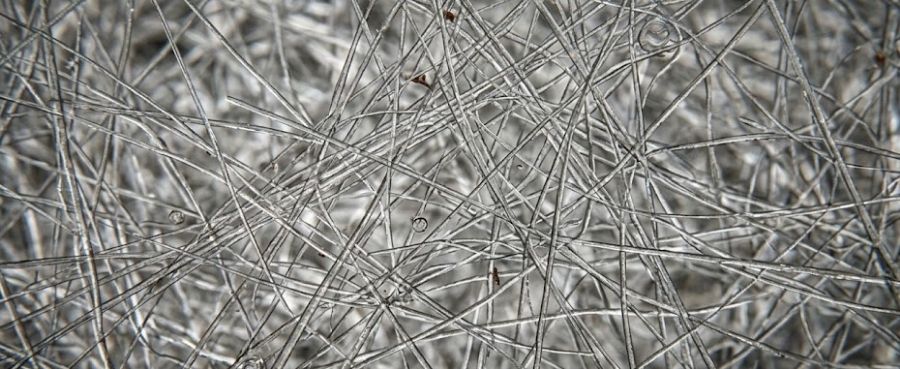
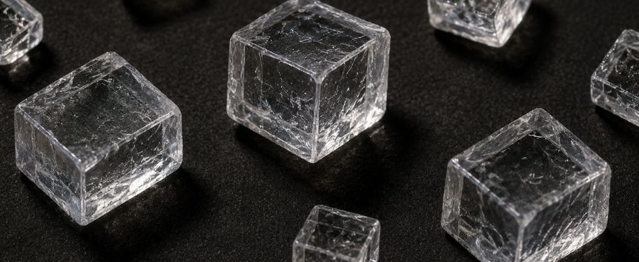
Before the microscope, humans believed that if something could not be seen, it simply did not exist. Magnification proved that our visible world is entirely built upon a hidden, microscopic foundation. It revealed the intricate cellular structure of plants, the microscopic crystalline composition of rocks, and the hidden fibers that make up synthetic and natural materials.
Changing Science and Medicine
In medicine, the microscope directly led to the germ theory of disease. Once scientists could actually see bacteria and pathogens, they could finally understand how illnesses spread and how to stop them. This tool enabled the creation of vaccines, the discovery of antibiotics, and the development of modern surgical pathology, ultimately saving countless millions of human lives.
How Modern Digital Microscopes Continue This Evolution
Today, magnification is no longer restricted to elite scientific laboratories. Digital technology has democratized microscopy, making it practical for everyday professional and hobbyist use.
Digital Microscopes Make Observation More Accessible
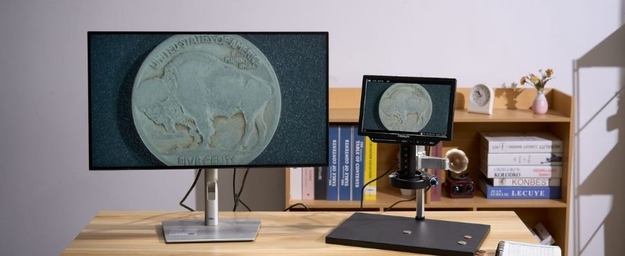
By removing the physical strain of looking through small eyepieces, digital models allow users to maintain a comfortable, upright posture while working. This accessibility is a game-changer for people performing repetitive industrial tasks, like quality control inspectors checking tiny welds, or hobbyists spending hours grading rare coins and inspecting stamps. The ability to instantly save and email high-resolution images has transformed global collaboration.
Choosing a Modern Microscope for Your Needs
When selecting a digital microscope today, your choice depends entirely on your daily tasks. If you need to solder electronics or repair jewelry, you require a model with a tall, stable stand that offers plenty of physical working distance for your tools. If you are a coin collector looking for mint errors, a model with built-in adjustable LED lighting and an integrated LCD screen provides the perfect portable setup for easy inspection.
A Simple Microscope History Timeline
First Compound Microscope (1590)
Zacharias and Hans Janssen construct the first sliding tube microscope, proving multi-lens magnification is possible.
Discovery of Cells (1665)
Robert Hooke publishes Micrographia and coins the biological term "cell" after observing cork wood.
First Microorganisms Seen (1674)
Antonie van Leeuwenhoek uses a highly refined single-lens microscope to discover living bacteria and protozoa.
First Electron Microscope (1931)
Ernst Ruska and Max Knoll break the light barrier, using electron beams to achieve extreme magnification.
First Digital Microscope (1986)
The digital era begins, replacing traditional eyepieces with camera sensors and monitors for collaborative viewing.
Final Thoughts
The evolution of the microscope from a simple arrangement of glass lenses to a highly advanced digital imaging system is a testament to human curiosity. Every major leap in optical technology—from discovering the first bacteria to inspecting modern microchips on a digital screen—has expanded our ability to understand and manipulate the world around us. Today's digital microscopes carry on this proud legacy, making microscopic exploration more comfortable, shareable, and accessible than ever before.
FAQs
What were the first things seen under a microscope?
The earliest users looked at common, everyday objects that were small but still visible to the naked eye. They primarily examined insects like fleas and flies, human hair, woven textiles, and the surface textures of wood and leaves. It was not until Leeuwenhoek's improved lenses that invisible microorganisms were finally seen.
Did Galileo Galilei invent the microscope?
No, Galileo did not invent the original concept, but he was an early pioneer. In 1609, he adapted his telescope design to focus on close objects rather than distant stars, creating his own version of a compound microscope called the occhiolino (little eye).
What is the most powerful microscope?
Transmission Electron Microscopes (TEM) are currently the most powerful type of microscope in existence. By passing a beam of electrons through ultra-thin specimens in a vacuum, they can magnify objects up to 50 million times, allowing scientists to see individual atoms.
What's the smallest thing we've ever seen?
Using advanced electron microscopy and scanning tunneling microscopes, scientists have been able to view and physically manipulate individual atoms. We can clearly see the atomic lattice structure of materials like gold and graphene, marking the absolute physical limit of current magnification technology.

Commenta
Nota che i commenti devono essere approvati prima di essere pubblicati.
Questo sito è protetto da hCaptcha e applica le Norme sulla privacy e i Termini di servizio di hCaptcha.