
Table of Contents
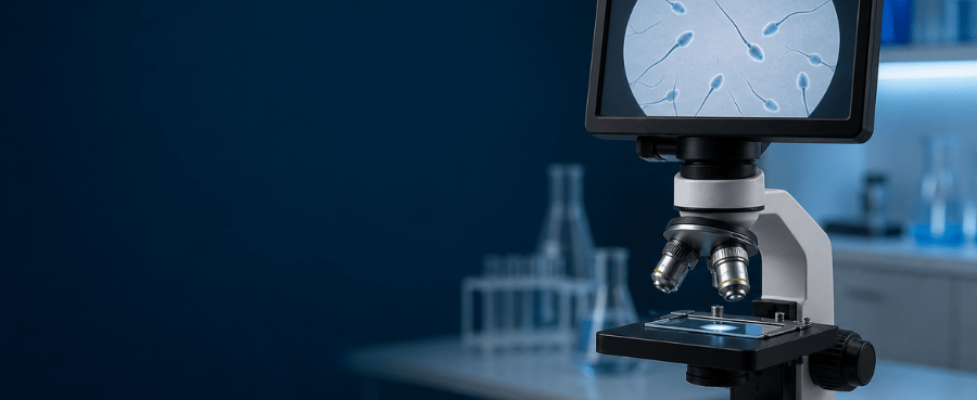
You may detect sperm as moving points at around 100x under favorable conditions, but approximately 200x to 400x total magnification is more practical for observing individual sperm cells and their movement. At 400x, the head and tail are generally easier to distinguish.
However, seeing sperm through a microscope is not the same as performing a semen analysis. A home microscope cannot reliably determine sperm concentration, total count, progressive motility, morphology or fertility. Detailed clinical morphology assessment typically requires a stained slide, trained interpretation and approximately 1000x total magnification with an oil-immersion objective.
This guide explains what you may see at each magnification, which type of microscope works best and why home observation should never replace professional testing.
Sperm Microscope Magnification at a Glance
The magnification you need depends on what you are trying to observe.
| Total magnification | What you may see | Best use | Main limitation |
|---|---|---|---|
| 40x | The overall sample area, fluid boundaries and larger debris | Locating the sample | Usually too low to identify individual sperm |
| 100x | Possible movement or tiny moving points under good conditions | Initial scanning | Head and tail are difficult to distinguish |
| 200x | Individual sperm and general movement become more practical to observe | General observation and some laboratory counting procedures | Fine morphology remains unclear |
| 400x | Individual sperm, heads, tails and movement are easier to see | Educational observation and closer examination | Cannot provide a complete fertility assessment |
| 1000x with oil immersion | Fine details in professionally prepared and stained smears | Laboratory morphology assessment | Requires staining, oil immersion and trained interpretation |
For most educational observations, 200x to 400x total magnification is the useful range. Higher magnification is not automatically better because image quality also depends on resolution, contrast, illumination and sample preparation.
If you are unfamiliar with objective and eyepiece numbers, see our guide to how microscope magnification is calculated.

What Does Total Magnification Mean?
On a traditional compound microscope, total magnification is calculated by multiplying the eyepiece magnification by the objective magnification:
Total magnification = Eyepiece magnification × Objective magnification
For example:
- 10x eyepiece × 4x objective = 40x total magnification
- 10x eyepiece × 10x objective = 100x total magnification
- 10x eyepiece × 20x objective = 200x total magnification
- 10x eyepiece × 40x objective = 400x total magnification
- 10x eyepiece × 100x objective = 1000x total magnification
This distinction matters because a “40x lens” may refer to a 40x objective rather than 40x total magnification. When combined with a 10x eyepiece, a 40x objective produces 400x total magnification.
Digital microscopes are more complicated. Their advertised magnification may depend on the lens, camera sensor, screen size and digital enlargement. A claimed 1000x digital image is therefore not necessarily equivalent to 1000x optical magnification through a compound microscope.
For a closer comparison of the common magnification levels, read 40x vs. 100x vs. 400x magnification.
Can You See Sperm at 40x Magnification?
At 40x total magnification, you can survey a relatively wide area of the slide, but individual human sperm cells are generally too small and transparent to identify reliably.
You may see:
- The edge of the liquid under the coverslip
- Air bubbles
- Fibers or dust
- Larger cells and debris
- General movement within the liquid
Movement at 40x should not automatically be interpreted as sperm. Fluid currents, debris and movement caused by pressure on the coverslip can create misleading results.
Use 40x mainly to locate the sample and bring the slide into focus before moving to a higher magnification.
Can You See Sperm at 100x Magnification?
At 100x total magnification, sperm may appear as tiny moving points if the microscope has good optics, suitable transmitted illumination and adequate contrast.
However, 100x usually does not provide enough detail to identify the head and tail clearly. It is better for scanning the sample than for examining individual cells.
At 100x, you may be able to:
- Detect possible movement
- Find areas that contain more moving cells
- Survey a wider field of view
- Center an area before switching to 200x or 400x
What you cannot reliably do at 100x is determine whether sperm concentration, motility or morphology is medically normal.
A moving point is not automatically a sperm cell, and the absence of visible movement does not prove that sperm are absent.
What Can You See at 200x Magnification?
At 200x total magnification, individual sperm become more practical to observe. With sufficient contrast, you may see their general shape and follow their movement across the field.
This magnification is commonly produced by a 10x eyepiece and a 20x objective.
At 200x, you may observe:
- Individual sperm cells
- General swimming movement
- Differences between moving and non-moving objects
- A larger field of view than at 400x
- More cells in the same field
The WHO Laboratory Manual for the Examination and Processing of Human Semen, Sixth Edition describes professional laboratory procedures that use phase-contrast optics at 200x or 400x for certain counting and assessment tasks.
That does not mean a home user can reproduce a clinical semen analysis simply by selecting the same magnification. Laboratories also use controlled sample depth, counting chambers, defined temperatures, standardized timing, replicate counts, quality-control procedures and trained interpretation.
What Can You See at 400x Magnification?
For most educational observations, 400x total magnification provides the most useful balance between detail, field of view and ease of focusing.
A standard compound microscope usually produces 400x total magnification with:
10x eyepiece × 40x objective = 400x total magnificationAt 400x, you may be able to see:
- Individual sperm cells
- The general outline of the head
- The tail
- Swimming movement
- Changes in direction
- Moving and apparently non-moving cells
- Debris and other cells in the sample
The 40x objective used to produce 400x total magnification is normally a dry objective. Immersion oil is not generally required at this level.
Although 400x provides a clearer view, it still cannot tell you whether a semen sample is clinically normal. A professional assessment considers multiple measurements, including sperm concentration, total sperm number, progressive and non-progressive motility, vitality and morphology.
These measurements cannot be estimated reliably from a casual view of one field.
Do You Need 1000x Magnification to See Sperm?
No. You do not normally need 1000x magnification merely to see sperm or observe movement.
A 1000x setup usually combines a 10x eyepiece with a 100x oil-immersion objective:
10x eyepiece × 100x oil-immersion objective = 1000x total magnificationProfessional laboratories may use approximately 1000x total magnification to evaluate sperm morphology on properly prepared and stained smears. This is different from observing live movement in an unstained sample.
According to the WHO laboratory manual, a stained morphology smear may first be surveyed at approximately 400x, while detailed assessment is performed with a 100x oil-immersion objective and at least a 10x eyepiece.
Using a 100x objective correctly requires:
- A suitable compound microscope
- An oil-immersion objective
- Immersion oil
- A properly fixed and stained smear
- Correct condenser settings
- Careful cleaning of the objective
- Training in morphology assessment
More magnification does not automatically reveal more useful detail. Once the optical system reaches its resolution limit, additional enlargement produces a larger but not necessarily clearer image.
What Type of Microscope Do You Need to See Sperm?
A compound transmitted-light microscope is generally the most suitable type for viewing sperm.
Unlike a surface-inspection microscope, transmitted illumination directs light upward through a thin sample on a glass slide. This is important because sperm are small, transparent cells rather than opaque surface objects.
A practical educational setup should include:
- A compound optical system
- Transmitted illumination
- A 10x eyepiece
- 10x, 20x and/or 40x objectives
- Fine-focus control
- An adjustable condenser or iris diaphragm
- A stable stage
- Standard glass slides and coverslips
Brightfield compound microscope
A standard brightfield compound microscope can be used for general observation, especially at 200x and 400x.
Because live sperm are nearly transparent, contrast may be limited. Lowering excessive brightness and carefully adjusting the condenser and iris diaphragm can make transparent objects easier to distinguish.
Phase-contrast microscope
Phase contrast is particularly useful for observing live, unstained sperm because it improves the visibility of transparent cells without requiring ordinary staining.
It is widely used in professional semen-analysis procedures, but it requires compatible phase objectives and a matching phase condenser. It is not simply a filter that can be added to every microscope.
Darkfield microscope
Darkfield illumination can make sperm appear bright against a dark background, which can make movement easier to see.
However, darkfield requires an appropriate condenser or optical configuration. It does not by itself turn a home observation into a standardized medical test.
Digital compound microscope
A digital compound microscope may work if it uses transmitted illumination and proper compound objectives. A screen can make movement easier to share, record and review.
The important word is compound. Many handheld and LCD digital microscopes are designed for reflective surface inspection rather than transparent biological slides.
If you are deciding between screen-based and eyepiece-based systems, read our digital vs. optical microscope comparison.
Handheld USB or LCD inspection microscope
A typical handheld USB microscope or LCD inspection microscope is usually designed for:
- Coins
- Circuit boards
- Jewelry
- Insects
- Textiles
- Mechanical surfaces
These devices commonly illuminate the sample from above. Even when the product advertises 500x, 1000x or more, it may lack the transmitted light, optical resolution, condenser and fine stage control needed for viewing transparent cells.
Do not choose a microscope for this purpose based only on the largest magnification number printed on the product page. Our guide to what to look for in a digital microscope explains why optical design and illumination matter more than exaggerated digital zoom claims.
Stereo microscope
A stereo or dissecting microscope normally operates at a much lower magnification and is intended for larger, solid objects.
It is excellent for insects, circuit boards and mechanical work, but it is generally not suitable for viewing individual sperm cells.
Features That Matter More Than Advertised Magnification
A microscope marked “2000x” is not automatically more useful than a well-designed 400x compound microscope.
Before selecting a microscope, check the following features.
Transmitted illumination
Light must pass through the sample. A ring light positioned above the slide is intended mainly for opaque surfaces and may not provide the necessary contrast.
Optical objectives
Look for clearly identified objective lenses. A 20x or 40x objective paired with a 10x eyepiece provides the most useful range for general observation.
Fine focus
At 400x, the depth of field is shallow. Small movements can take the sample out of focus, so a fine-focus control is important.
Condenser and iris diaphragm
These controls help direct and shape the light passing through the sample. They can improve contrast when viewing nearly transparent cells.
Mechanical stability
A weak stand or unstable stage makes high-magnification focusing difficult. The sample should remain still while the focus and stage position are adjusted.
Real resolution
Optical resolution determines whether two nearby details can be distinguished. Digital zoom only enlarges the pixels already captured and cannot restore missing detail.
Image recording
A camera is helpful for recording educational observations, but camera resolution does not replace optical resolution. The optical image must be clear before a higher-resolution sensor can provide a useful recording.
Seeing Sperm Is Not the Same as Performing a Semen Analysis

This distinction is the most important part of home observation.
A microscope may allow you to see objects that appear to be sperm, but a clinical semen analysis requires much more than visual confirmation.
| A home observation may show | A home observation cannot reliably determine |
|---|---|
| Possible sperm-like cells | Accurate sperm concentration |
| Whether some cells appear to move | Total sperm number |
| General movement in one field | Percentage of progressive motility |
| A basic head-and-tail outline | Percentage of normal morphology |
| An educational view of the sample | Fertility or infertility |
| A video of one selected area | Whether a vasectomy was successful |
A single field of view may not represent the entire sample. The WHO manual specifically notes that the absence of sperm from an examined portion does not necessarily mean sperm are absent from the remainder of the ejaculate.
Clinical laboratories reduce this uncertainty through standardized mixing, calibrated counting chambers, replicate measurements and quality-control procedures.
Can home observation assess sperm health?
No. Terms such as “healthy sperm” involve several laboratory measurements and clinical interpretation. Seeing cells swim does not establish that their concentration, progressive motility, morphology or genetic quality is normal.
Can home observation confirm fertility?
No. Fertility depends on both partners and cannot be confirmed by looking at one semen sample. Even a professional semen analysis does not independently prove that an individual or couple is fertile.
Can a microscope confirm a successful vasectomy?
No. Do not use a home microscope to decide whether contraception is no longer required after a vasectomy.
Post-vasectomy clearance should be based on the testing schedule and laboratory result provided by the treating clinician. Failure to see sperm at home is not reliable evidence of sterility.
Why Can’t I See Sperm Under My Microscope?
Several technical and biological factors can make observation difficult.
You are using the wrong type of microscope
A stereo microscope or reflective-light inspection microscope may not be able to produce a useful image of a transparent slide.
Use a compound transmitted-light microscope rather than relying only on an advertised digital magnification number.
You started at too high a magnification
A high-power objective has a narrow field of view and shallow depth of field. Begin at low power to locate and center the sample before switching to 200x or 400x.
At 400x, use only the fine-focus control. Moving the stage too far with coarse focus can cause the objective to contact the slide.
For general focusing principles, see how to use a digital microscope. Keep in mind that biological slides require transmitted illumination and may differ from the opaque samples shown in that guide.
The illumination is too bright
Excessive brightness can wash out transparent cells. Adjust the condenser and partially close the iris diaphragm to increase contrast.
Do not reduce the light so much that the image becomes grainy or difficult to focus.
The microscope lacks optical resolution
A large number on the screen does not guarantee useful detail. Cheap digital zoom may enlarge a blurry image without resolving the head or tail.
The sample layer is difficult to focus through
A thick or uneven liquid layer, air bubbles, fingerprints, fibers and debris can interfere with focusing. Multiple objects may also be present at different depths.
Movement is being confused with fluid motion
Liquid movement, pressure on the coverslip and temperature changes can move debris through the field. Motion alone does not prove that an object is a sperm cell.
No sperm are visible in the area examined
The absence of visible sperm in one field or one small portion cannot be interpreted as proof that none are present in the entire sample.
If the question relates to fertility, post-vasectomy clearance or another medical decision, use an accredited laboratory or consult a qualified healthcare professional.
Do You Need to Stain Sperm to See Them?
Staining is not normally required simply to observe live sperm movement. In fact, the preparation process used for fixed and stained smears means the cells are no longer being observed for live motility.
Professional morphology assessment is different. Laboratories prepare, fix and stain a smear so trained personnel can examine the proportions and structures of sperm cells under controlled conditions, commonly using oil immersion at approximately 1000x total magnification.
Do not assume that a colored household substance or an improvised stain is appropriate. Laboratory stains and preparation methods are standardized, and some reagents require specific handling precautions.
Can a Phone Microscope See Sperm?
A simple clip-on phone lens is unlikely to provide the combination of optical resolution, transmitted illumination, contrast and mechanical control needed for reliable viewing.
A phone attached to a suitable compound microscope may record what the microscope already resolves. In that case, the compound microscope supplies the important optical image, while the phone functions mainly as a camera.
The phone’s digital zoom does not increase the microscope’s resolving power.
Can a USB Microscope See Sperm?
Some digital compound microscopes with transmitted illumination may show sperm. Most handheld USB surface-inspection microscopes are less suitable because they are optimized for opaque objects illuminated from above.
Before using a USB model, verify that it has:
- True transmitted illumination
- A slide stage
- Sufficient optical resolution
- Stable fine focusing
- A suitable field of view
- Real sample images captured from transparent biological slides
Do not rely on advertised magnification alone.
Basic Hygiene and Safe Handling
Human semen is a body fluid and should be handled carefully.
For educational handling:
- Wash your hands before and after handling the sample.
- Cover cuts or broken skin.
- Avoid contact with the eyes, mouth and shared surfaces.
- Do not eat or drink in the work area.
- Use clean slides and coverslips.
- Do not share materials that have contacted a sample.
- Clean the work surface after observation.
- Dispose of contaminated single-use materials appropriately.
- Follow the microscope manufacturer’s instructions when cleaning the stage and lenses.
Do not place alcohol, household cleaner or water directly on an objective lens. Use appropriate lens paper and manufacturer-approved optical cleaning methods.
If the sample belongs to another person, professional biosafety and consent considerations apply. Home observation is not an appropriate substitute for controlled laboratory handling.
Frequently Asked Questions
What magnification do you need to see sperm?
Approximately 200x to 400x total magnification is the most practical range for observing individual sperm and movement. At 100x, movement may be detectable, but individual cells are harder to identify clearly.
Can you see sperm at 100x?
Possibly. With good optics and contrast, sperm may appear as tiny moving points. However, 100x is better used for scanning than for identifying individual heads and tails.
Is 200x enough to see sperm?
Yes, 200x may be sufficient for observing individual sperm and general movement when the microscope has suitable optics, transmitted illumination and contrast. Professional laboratory procedures may use phase contrast at 200x or 400x for certain assessments.
Can you see sperm clearly at 400x?
At 400x, individual sperm, their general head-and-tail outline and movement are usually easier to observe. “Clearly” does not mean that clinical concentration, motility or morphology can be determined from a home observation.
What objective lens gives 400x total magnification?
A 40x objective combined with a 10x eyepiece gives 400x total magnification. This is normally a dry objective and does not usually require immersion oil.
Is 1000x necessary to see sperm?
No. It is not necessary merely to see sperm or observe movement. Approximately 1000x with an oil-immersion objective is used for detailed professional assessment of properly prepared and stained morphology smears.
Do you need immersion oil at 400x?
No. A standard 40x objective producing 400x total magnification is normally used dry. Immersion oil is typically used with a 100x oil-immersion objective to produce approximately 1000x total magnification.
Do sperm need to be stained?
Not for observing live movement. Staining is generally associated with fixed preparations used for professional morphology assessment.
Can a digital microscope see sperm?
A digital compound microscope may work if it has transmitted illumination, suitable compound objectives and adequate optical resolution. A typical handheld digital inspection microscope may not.
Why can’t I see sperm at 400x?
Possible causes include incorrect illumination, insufficient contrast, poor focus, an unsuitable microscope design, a thick or uneven sample, optical limitations or examining an unrepresentative area. The absence of visible sperm at home should not be interpreted medically.
Can a home microscope test male fertility?
No. It cannot reliably measure all the parameters required for a professional semen analysis, and it cannot diagnose fertility or infertility.
Can I use a home microscope after a vasectomy?
A home microscope must not be used to confirm post-vasectomy clearance. Continue following the clinician’s contraception and laboratory-testing instructions until formal clearance is provided.
Final Answer
For general educational observation, choose a compound transmitted-light microscope capable of approximately 200x to 400x total magnification. A 10x eyepiece with a 20x or 40x objective is a practical configuration.
At 100x, you may detect movement. At 200x, individual sperm become easier to observe. At 400x, the head, tail and movement are generally clearer. Approximately 1000x with oil immersion is mainly associated with professional examination of prepared and stained morphology smears—not ordinary live observation.
Most importantly, seeing sperm through a microscope does not measure fertility, confirm normal sperm quality or replace a professional semen analysis.

发表评论
所有评论在发布前都会经过审核。
此站点受 hCaptcha 保护,并且 hCaptcha 隐私政策和服务条款适用。