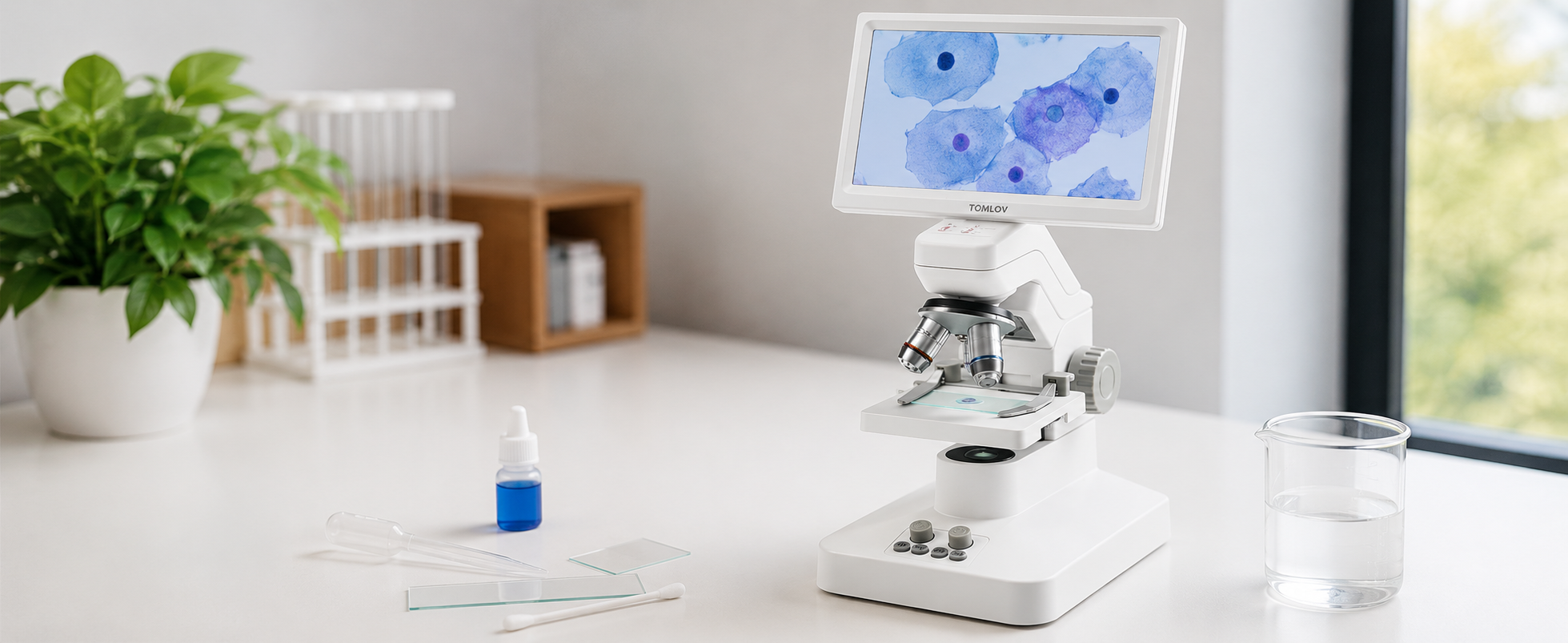
Cheek Cells Under a Microscope: A Beginner’s Wet Mount Guide
Learn how to prepare and view cheek cells under a microscope using a wet mount, methylene blue stain, bottom lighting, and beginner-safe steps.
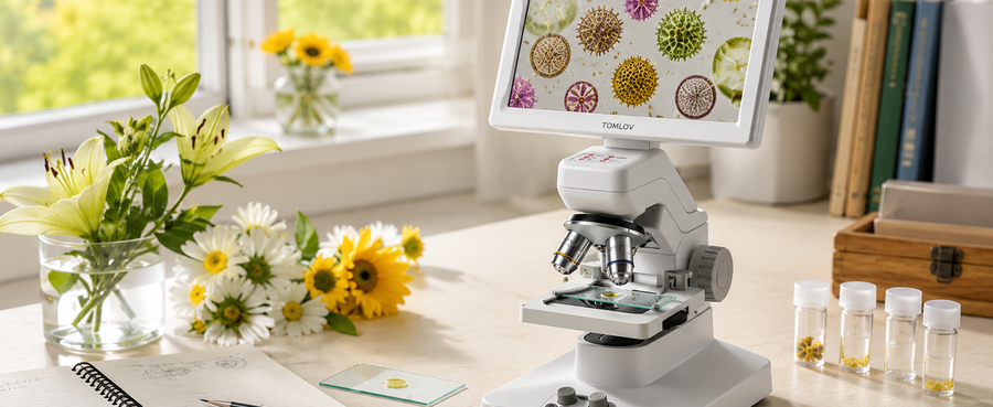
Pollen Under the Microscope: A Beginner’s Step-by-Step Observation Guide
Pollen grains appear round, oval, spiky, ridged, or clustered under a digital microscope, with tips for dry samples, wet mounts, and lighting.
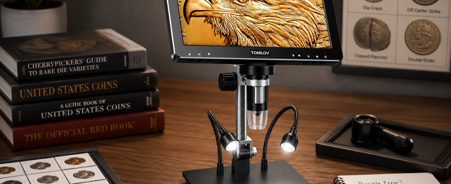
Mint Error vs. Post-Mint Damage: What to Check Under a Coin Microscope
Mint errors form during striking, while post-mint damage happens later; learn what to check on rims, marks, surfaces, and coin details.
Parts of a Microscope and Their Functions: A Complete Beginner’s Guide
Learn the main parts of a microscope, what each part does, and how traditional and digital microscope components differ.
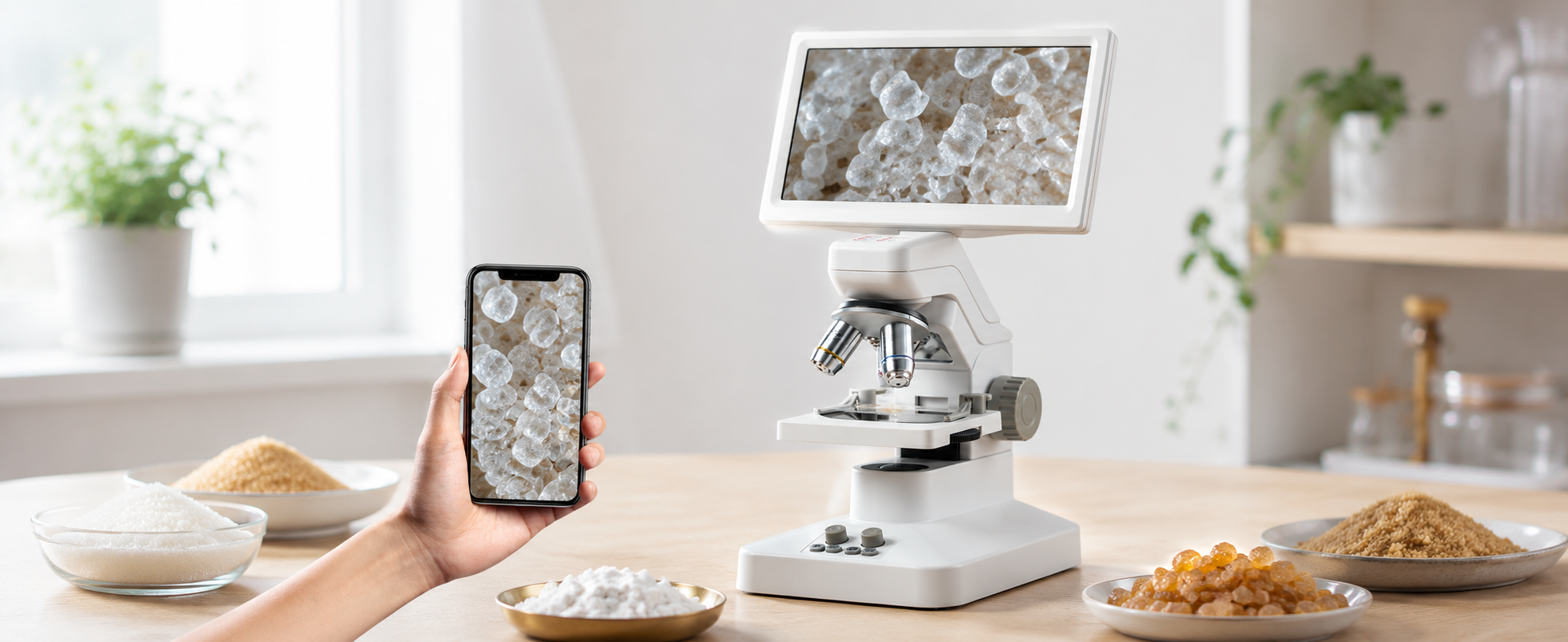
Sugar Crystals Under a Digital Microscope: A Beginner’s Observation Guide
See how granulated, brown, powdered, and rock sugar look under a digital microscope, with setup, lighting, focus, and comparison tips.
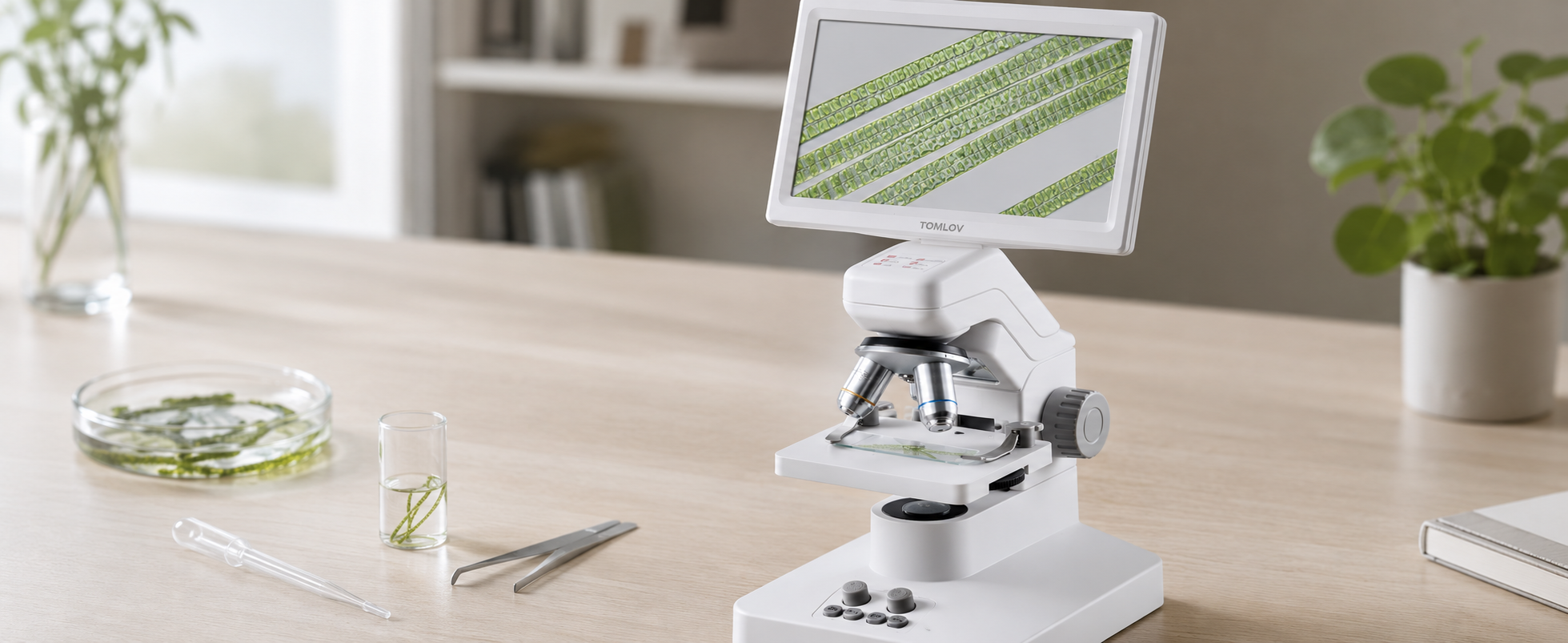
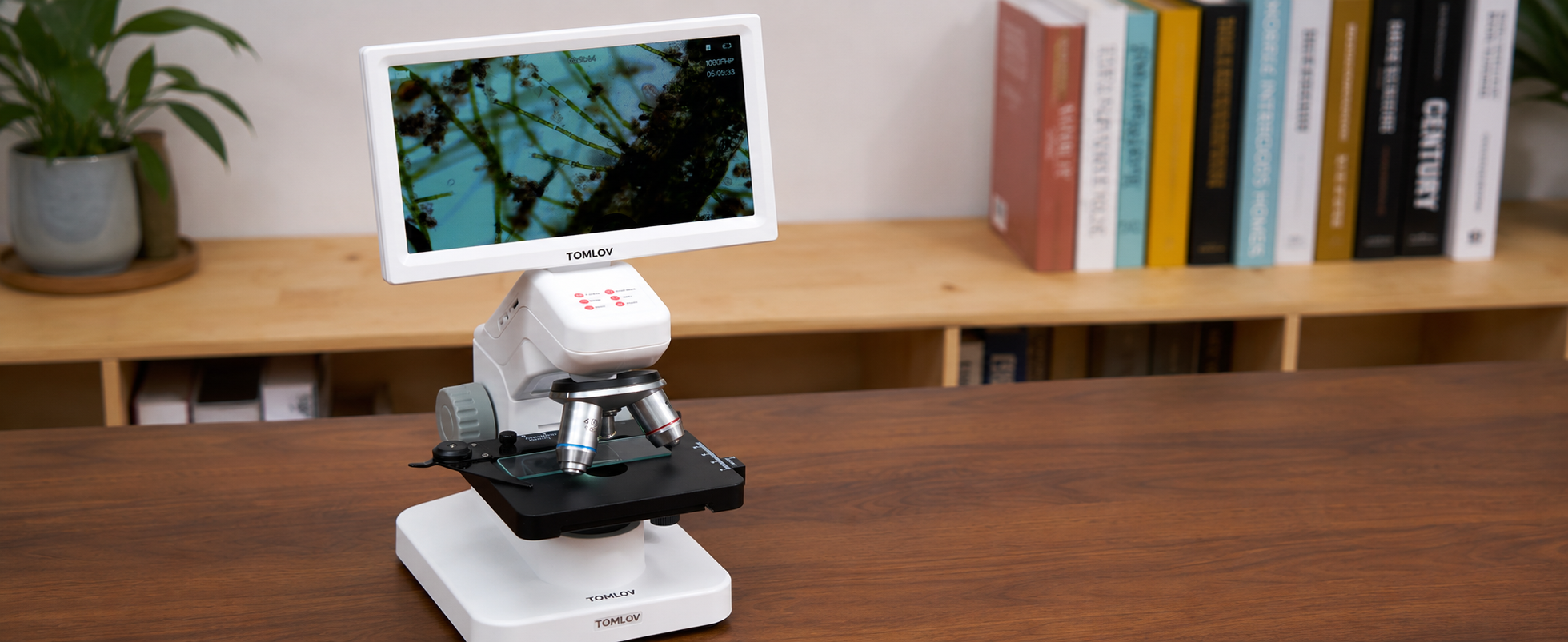
Spirogyra Under a Microscope: How to Prepare and Observe a Wet Mount
Learn how to prepare a Spirogyra wet mount, adjust magnification, and identify spiral chloroplasts, cell walls, and connected filaments.
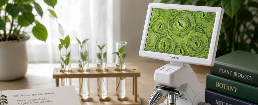
Plant Stomata Under a Microscope: How to Prepare and Observe a Leaf Slide
Apply clear nail polish to a leaf, lift the dry impression with tape, and place it on a slide. View under a microscope to observe plant stomata and guard cells.
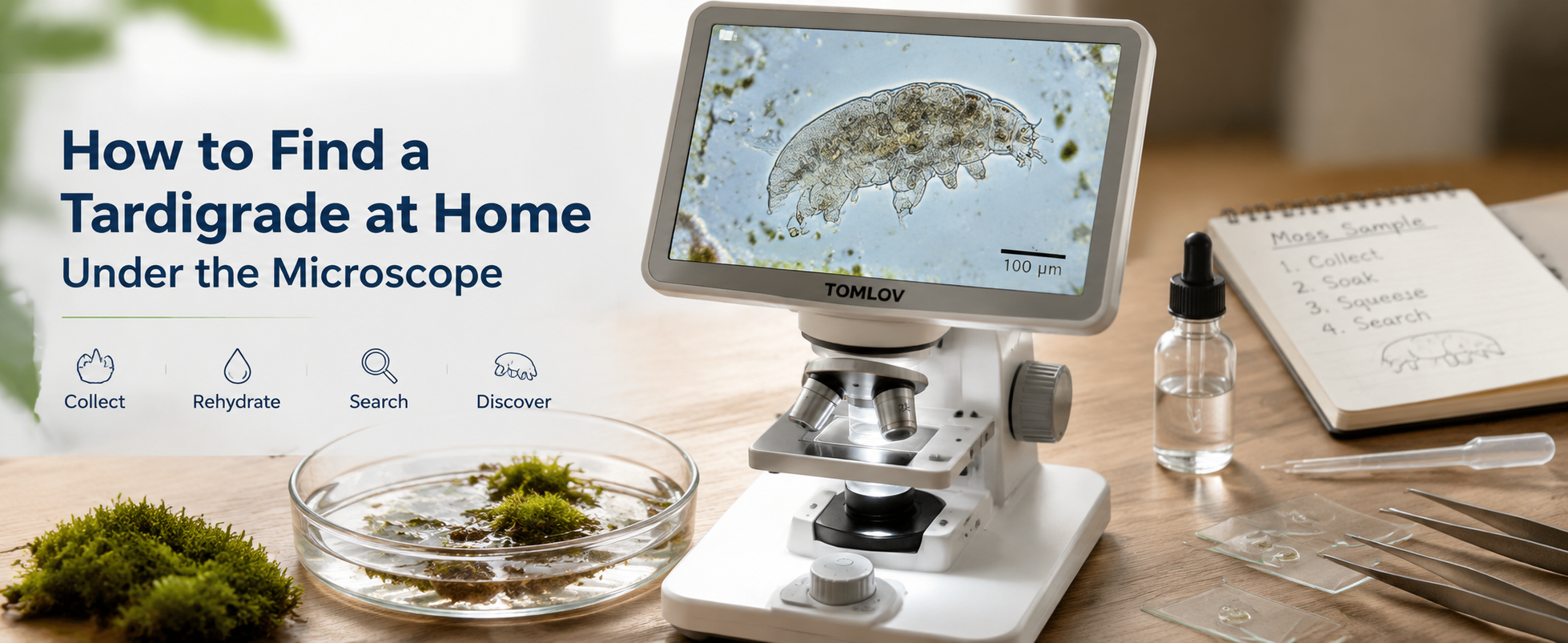
How to Find a Tardigrade at Home Under the Microscope
Find tardigrades at home by soaking moss or lichen, squeezing out the water, and scanning small drops under a microscope; then transfer one for closer viewing.
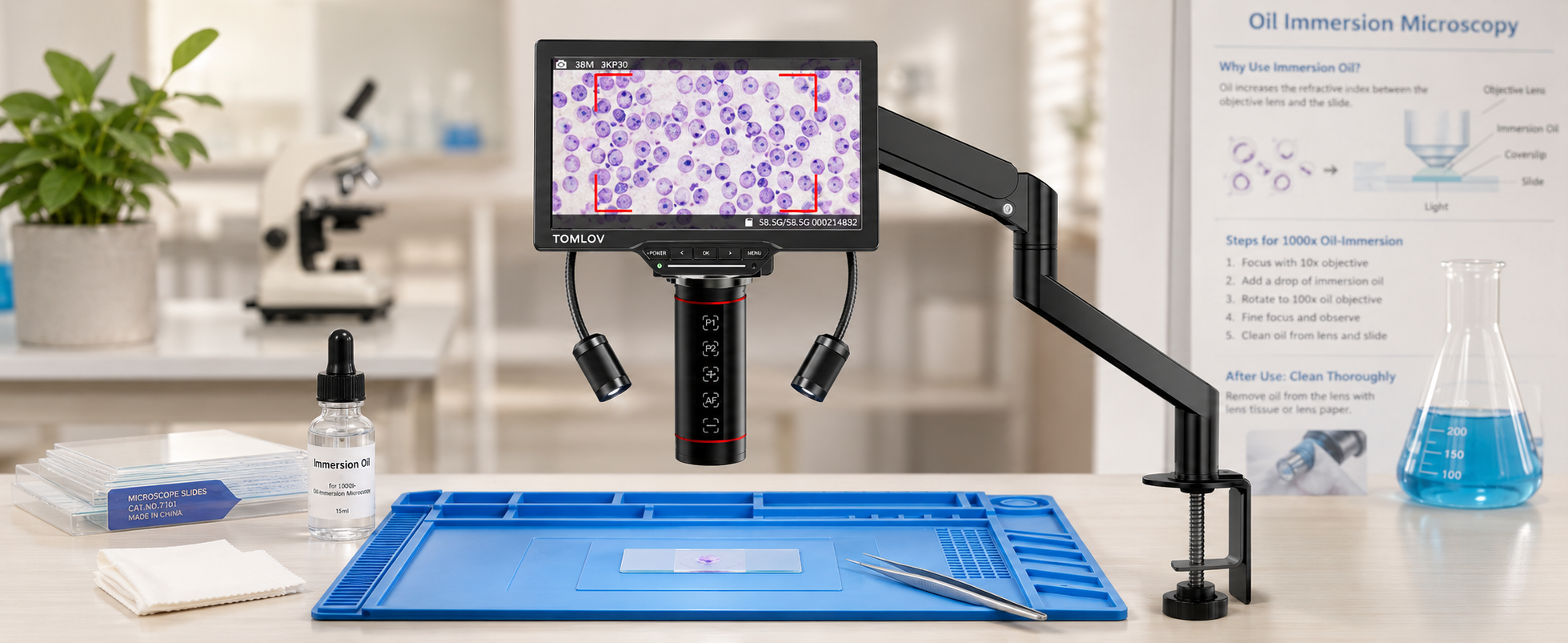
How to Use Microscope Immersion Oil: When and Why It's Needed
Learn when and why to use microscope immersion oil with a 100x objective, how it improves resolution, and how to apply, focus, and clean it safely.