
Table of Contents
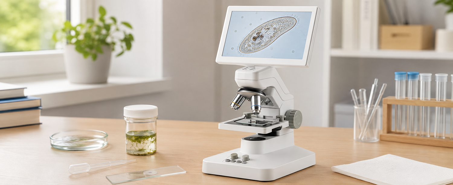
Paramecium is a fast-moving single-celled organism often found in pond water or prepared cultures. Under a microscope, it usually looks like a tiny slipper-shaped body moving through water with the help of cilia. To view it clearly, you need a thin wet mount, transmitted light from below, moderate magnification, and a way to slow the organism without damaging it.
What Does Paramecium Look Like Under a Microscope?
A Slipper-Shaped Single-Celled Organism
Paramecium is often described as slipper-shaped because its body is long, rounded, and slightly tapered. Under low magnification, it may first appear as a tiny moving speck. Once you focus more carefully, the outline becomes easier to recognize.
The body is translucent, so it will not look strongly colored unless the lighting and contrast are adjusted well.
Fast Movement Through Water
One of the first things beginners notice is how fast Paramecium moves. It may swim across the field of view before you can focus on it. This is normal.
Because of that movement, start at low magnification and follow the organism before switching to a closer view. A crowded or deep water drop makes tracking even harder.
Cilia, Oral Groove, and Visible Internal Structures
Paramecium moves with tiny hair-like structures called cilia. At beginner magnification, cilia may appear as a faint shimmering edge rather than individual hairs. You may also see the oral groove along one side of the body and darker internal areas such as food vacuoles.
These details are easiest to notice when the organism slows down and stays in focus.

What Should You Prepare Before Viewing Paramecium?
Pond Water, Infusion Water, or Prepared Paramecium Culture
You can observe Paramecium from pond water, infusion water, or a prepared culture from an educational supplier. Pond samples may also contain algae, debris, and other microorganisms, so they can be interesting but less predictable.
Infusion water, often made from plant material in water, may encourage microorganisms to grow. Use clean containers and avoid unsafe or polluted water sources.
A Microscope With Transmitted Light and 100× to 400× Magnification
Paramecium is transparent, so transmitted light from below the slide is important. A top-light-only magnifier is not ideal for this wet-mount sample.
For beginner observation, a digital microscope for specimen observation should support slide viewing with bottom light, not just surface inspection. Around 100× to 400× total magnification is usually enough for shape, movement, and basic internal details.
Slides, Cover Slips, Droppers, and Paper Towels
Prepare clean glass slides, cover slips, a dropper or Pasteur pipette, paper towels, and a small container of sample water. Clean glass matters because dust and grease can look confusing under magnification.
Paper towels are useful for absorbing extra water around the cover slip so the sample does not flow too much.
Optional Slowing Methods: Methyl Cellulose or Cotton Fibers
Paramecium can move too quickly for beginners to follow. A tiny amount of methyl cellulose, sometimes sold for slowing protozoa, can thicken the water and reduce movement.
A few clean cotton fibers can also help by creating a light barrier that slows swimming. Use very little. Too much can crowd the slide and block the view.
How Do You Prepare and View a Paramecium Wet Mount?
Collect Sample Water From Cloudy or Organic-Rich Areas
Paramecia often feed near bacteria-rich areas, so the best sample may come from slightly cloudy water, the lower part of a culture jar, or near decaying plant material. Draw only a small amount with a dropper.
Do not stir the whole container aggressively. Gentle sampling keeps the organisms and debris easier to manage.
Place One Small Drop in the Center of the Slide
Place one small drop of sample water in the center of a clean slide. A large drop may look helpful, but it usually makes the organism drift too much.
A thin wet mount is easier to focus. It also keeps Paramecium closer to one focus plane, which makes tracking and recording much easier.
Add a Slowing Agent if the Paramecia Move Too Fast
If the Paramecia move too quickly, add a very small amount of methyl cellulose or introduce a few cotton fibers near the drop. The goal is to slow movement, not trap or crush the organism.
Do not overload the slide. Too much slowing agent can make the sample cloudy or prevent clear observation.
Lower the Cover Slip at a 45-Degree Angle
Hold the cover slip at about a 45-degree angle. Touch one edge to the liquid first, then lower it slowly. This helps push air outward and reduces large bubbles.
A few small bubbles are normal, but large bubbles can block the view and move the sample around.
Remove Excess Water With Tissue Paper
If water leaks from the cover slip edges, gently touch the edge with tissue paper. The tissue will draw away extra liquid without disturbing the whole slide.
Do not press down on the cover slip. Pressing can squeeze the sample, move the organism suddenly, or damage the wet mount.
Start at Low Magnification to Find Moving Organisms
Begin with the lowest objective or a low total magnification. Paramecium is large enough to spot as a fast-moving shape before you zoom in.
Use slow slide movements and scan the whole drop. Starting too close makes it easy to miss the organism completely.
Adjust Bottom Light and Focus for Better Contrast
Paramecium is translucent, so too much light can make it disappear. Reduce the bottom light slightly or adjust the diaphragm if your microscope has one. Lower light often improves contrast and makes the body outline easier to see.
For moving wet mounts, the TOMLOV DM301 Pro digital microscope can support this workflow because beginners can adjust bottom light and focus while watching the live image on a screen.
Increase Magnification After Finding a Clear Paramecium
After you find a clear Paramecium, move to higher magnification gradually. Around 100× can show body shape and movement. Around 400× may help reveal cilia movement, vacuoles, and the oral groove more clearly.
Refocus after each magnification change. Do not expect every structure to be visible in every sample.
Save Images or Videos Before the Sample Dries
Wet mounts can dry, shift, or change quickly. Save images or short videos while the organism is still moving and visible. A short video can be more useful than a single photo because Paramecium is active.
For students documenting observations, a 4K digital microscope can help keep clearer visual records for notes, comparisons, or lab reports.

What Details Should Beginners Look For?
Body Shape and Swimming Direction
Start by watching the body shape and movement. Paramecium usually swims with a smooth, gliding motion, although it may turn, reverse, or change direction when it meets debris.
Its slipper-like outline is one of the easiest beginner clues, especially when compared with rounder microorganisms or random particles.
Cilia Along the Cell Edge
Cilia are tiny structures along the outer edge. Under a beginner microscope, they may not appear as individual hairs. Instead, you may notice a shimmering fringe or fuzzy movement around the body.
This effect is easier to see when the light is not too bright and the organism is not swimming too quickly.
The Oral Groove on One Side of the Cell
The oral groove is a shallow indentation along one side of the Paramecium. Cilia near this groove help move food particles toward the cell mouth.
Beginners may not see it clearly at first. Look for a slightly indented side or an area where particles seem to move toward the cell.
Food Vacuoles and Contractile Vacuoles
Food vacuoles may appear as small darker circles inside the cytoplasm. Contractile vacuoles may look like clearer circular or star-like areas that fill and empty as the organism manages excess water.
These structures are not always easy to see. Focus first on the body outline and movement, then look for internal details.
Debris, Bubbles, and Other Microorganisms Nearby
Pond or culture samples often contain debris, bacteria-rich particles, algae, bubbles, and other microorganisms. Air bubbles usually have thick dark borders and do not swim. Debris may drift or sit still.
Do not assume every moving organism is Paramecium. Compare shape, movement, and visible internal features before labeling it.
What Mistakes Should Beginners Avoid?
Do Not Use Too Much Water on the Slide
Too much water makes Paramecium swim in a deeper space, which makes focusing harder. It can also cause the cover slip to float.
Use one small drop first. A thin wet mount gives you better control and keeps the organism easier to follow.
Do Not Start at the Highest Magnification
High magnification gives a narrow field of view. If you start too high, Paramecium may swim out of view immediately.
Start low, locate a moving organism, center it, and then increase magnification. This order saves time and frustration.
Do Not Use Too Much Slowing Agent
A slowing agent can help, but too much can make the sample thick, cloudy, or crowded. Cotton fibers can also block the view if you add too many.
Use the smallest amount possible. The goal is slower movement, not a trapped or unreadable sample.
Do Not Confuse Paramecium With Other Pond Microorganisms
Many pond organisms move under a microscope. Some are round, some are worm-like, and some dart quickly through the water. Paramecium is usually recognized by its slipper-like shape, cilia-driven movement, and translucent body.
Use those clues together rather than relying on one feature.
Do Not Let the Wet Mount Dry Out
As the slide dries, movement slows, focus changes, and organisms may become stressed or distorted. If the slide begins drying, add a tiny amount of water near the cover slip edge.
Do not flood the slide. A small addition is enough.
Do Not Treat Beginner Observation as Species Identification
A beginner wet mount can help you recognize Paramecium-like organisms, but it should not be treated as exact species identification. There are different Paramecium species, and proper identification may require more careful comparison.
Describe what you observe rather than making a final classification.
Conclusion
Viewing Paramecium under a microscope is a useful beginner wet-mount activity because it shows movement, cell shape, cilia-driven swimming, and internal structures in a living single-celled organism. The best results come from a small sample drop, a gently lowered cover slip, transmitted light, low magnification first, and careful focus adjustment. Slow the organism only if needed, save images or video before the slide dries, and avoid confusing Paramecium with bubbles, debris, or other microorganisms.
FAQs
What Magnification Is Needed to See Paramecium?
Paramecium can often be found at low magnification, but more detail appears around 100× to 400× total magnification. Start low to locate moving organisms, then increase magnification gradually. Clear transmitted light, a thin wet mount, and slower movement are often more important than using the highest magnification immediately.
What Is the Size of a Paramecium Under a Microscope?
Paramecium is usually large compared with many microorganisms, often around a few hundred micrometers long depending on the species. Under the microscope, it may first appear as a tiny moving speck, then become a slipper-shaped cell as you increase magnification and improve focus.
Is a Paramecium a Bacteria or Fungi?
Paramecium is neither bacteria nor fungi. It is a single-celled eukaryotic organism often classified as a ciliate protozoan. Unlike bacteria, it has more complex internal structures. Unlike fungi, it does not grow as mold or yeast. It moves through water using cilia.
Can Paramecia Be Seen Without a Microscope?
Individual Paramecia are too small to see clearly without a microscope. You may notice cloudy culture water or tiny moving specks under strong lighting, but you cannot observe their shape, cilia, or internal structures with the naked eye. A microscope is needed for meaningful observation.

发表评论
所有评论在发布前都会经过审核。
此站点受 hCaptcha 保护,并且 hCaptcha 隐私政策和服务条款适用。