
Table of Contents
Microscope magnification is how many times larger an object appears under the lens compared to the naked eye. A 100x view means the specimen looks one hundred times its actual size. The total figure comes from multiplying the eyepiece by the objective — and while that sounds simple, choosing the right combination is where most beginners stumble. This guide walks through the numbers, the standard lens ranges, and how to apply them in practice.
What is microscope magnification?
Magnification refers to the degree of visual enlargement produced by a microscope's lens system. It is expressed as a multiplier — 10x, 40x, 400x — indicating how many times larger the image appears than the actual specimen. A 40x view of a human hair, for example, shows it at the apparent thickness of a pencil.
Magnification alone, however, does not determine usefulness. A blurry image at 1000x reveals less than a sharp one at 100x. That is why magnification is best understood alongside resolution, which is covered later in this guide.
How do you calculate total magnification?
Total magnification is the product of two lenses working together: the eyepiece and the objective. Knowing how each contributes makes the math straightforward.
The role of the eyepiece
The eyepiece, or ocular lens, is the lens you look through at the top of the microscope. Most standard eyepieces are rated at 10x, though 15x, 20x, and even 30x versions exist. The eyepiece magnifies the image already produced by the objective below it.
The role of the objective lens
The objective lens sits closest to the specimen and does the primary work of magnification. Compound microscopes typically carry three or four objectives on a rotating nosepiece — commonly 4x, 10x, 40x, and 100x. The number engraved on the side of each objective tells you its power.
Multiplying the two together
Total magnification equals eyepiece × objective. A 10x eyepiece paired with a 40x objective yields 400x. Pair the same eyepiece with the 100x oil immersion lens and the total rises to 1000x. Digital instruments such as a digital microscope work differently — without an eyepiece, total magnification is determined by the lens, the sensor, and the display size combined.

What can you see at each magnification level?
Scanning power (40x total)
The 4x objective produces 40x total magnification. It is used to locate the specimen, survey the slide, and assess overall sample density. Pond water debris, plant tissue sections, and insect parts are clearly visible at this level.
Low power (100x total)
The 10x objective gives 100x total. This range reveals general tissue structure — leaf cell arrangements, the outlines of larger protozoa, and the body shape of small invertebrates such as rotifers.
High power (400x total)
The 40x objective yields 400x total magnification. Individual animal and plant cells, larger bacteria, and blood smear components become resolvable. This is the working range for most cellular biology observations.
Oil immersion (1000x total)
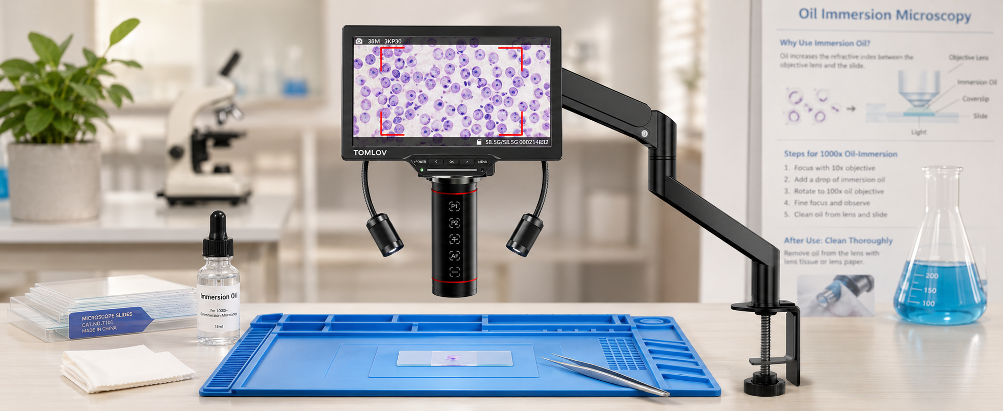
The 100x objective requires a drop of immersion oil between the lens and the coverslip to maintain resolution. Total magnification reaches 1000x, sufficient to distinguish microscopic bacteria and fine cellular detail. Without the oil, the image at this power loses clarity.
What magnification can different microscopes reach?
Stereo microscope
Stereo microscopes operate between roughly 2x and 90x, with some configurations reaching 180x using a Barlow lens. They produce a three-dimensional view at lower magnification and are suited to solid, opaque objects — circuit boards, coins, insects, and dissection work. A coin microscope typically falls within this category, offering enough magnification to inspect mint marks and surface wear without obscuring the full field.
Compound microscope
Compound light microscopes cover 40x to 1000x, occasionally extending to 1500x with specialized optics. They are designed for transparent specimens mounted on slides — cells, bacteria, water organisms, and thin tissue sections. This is the standard instrument in classrooms and home labs.
Electron microscope
Electron microscopes use electron beams rather than light and can reach 200,000x or higher. They resolve structures at the molecular level but are restricted to research facilities due to their size, cost, and operational complexity.

What other factors affect image quality?
Resolution and image clarity
Resolution is the ability to distinguish two adjacent points as separate. Light microscopes are limited by the wavelength of visible light to about 0.2 micrometers, which is why optical magnification beyond 1500x produces no additional usable detail.
Light and contrast
Translucent specimens require controlled illumination. Excessive light washes out detail, while insufficient light renders the image dim. The diaphragm beneath the stage adjusts contrast by narrowing or widening the light cone.
Field of view and working distance
As magnification increases, the visible area shrinks and the lens moves closer to the slide. At 1000x, the field of view is narrow and the working distance is less than a millimeter — which is why coarse focus must never be used at high power.
How should beginners pick the right magnification?
Start low and work your way up
Always begin at 40x. Lower magnification provides a wider field of view, making it easier to locate the specimen and assess what is on the slide. Once the target is centered, switch to 100x, then 400x, using the fine focus knob only.
Match the magnification to your sample
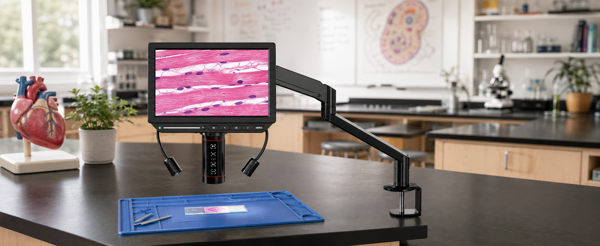
Larger structures — insects, plant parts, fabric weaves — are best viewed between 20x and 100x. Cellular detail requires 400x. Bacteria and subcellular features call for 1000x with oil immersion. For users who prefer screen-based viewing over an eyepiece, the Tomlov DM9 provides a 10.1-inch HDMI display suitable for the 40x–500x range covered in most beginner work.
Conclusion
Microscope magnification is a matter of multiplication and matching — eyepiece times objective, then the right total for the specimen at hand. The numbers themselves are simple. What takes practice is recognizing when a higher figure no longer adds detail and when image quality is being limited by light, resolution, or technique rather than power. Start at 40x, increase gradually, and treat magnification as one tool among several rather than the only one that matters.
FAQs
What can you see with 10x, 40x, and 100x objective lenses?
These three objectives cover the standard working range of a compound microscope. Paired with a 10x eyepiece, they yield 100x, 400x, and 1000x total magnification — used respectively for tissue structure, individual cells, and bacteria or fine cellular detail.
What is 40x, 100x, and 400x magnification?
These are the total magnifications produced by a 10x eyepiece combined with the 4x, 10x, and 40x objectives. 40x is for locating the specimen, 100x for general tissue structure, and 400x for individual cells and larger bacteria.
What does 200x magnification mean?
200x means the specimen appears two hundred times its actual size. On a compound microscope, this is typically achieved with a 10x eyepiece and a 20x objective, or a 20x eyepiece with a 10x objective. It sits between low and high power, useful for medium-detail observation.
Why does my image blur when I switch to a higher objective?
The most common cause is using the coarse focus knob at high power, which moves the stage too far in a single turn. At 400x and above, use the fine focus knob only. A persistent blur after fine focusing usually indicates a dirty lens, a thick or uneven sample, or focus set on debris in a different plane from the target.

Hinterlasse einen Kommentar
Alle Kommentare werden vor der Veröffentlichung geprüft.
Diese Website ist durch hCaptcha geschützt und es gelten die allgemeinen Geschäftsbedingungen und Datenschutzbestimmungen von hCaptcha.