
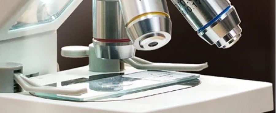
Microscope Stage Height Adjustment: How It Affects Focus
Learn how microscope stage height directly controls focus and working distance. Get step-by-step tips to adjust your stage safely for any sample.
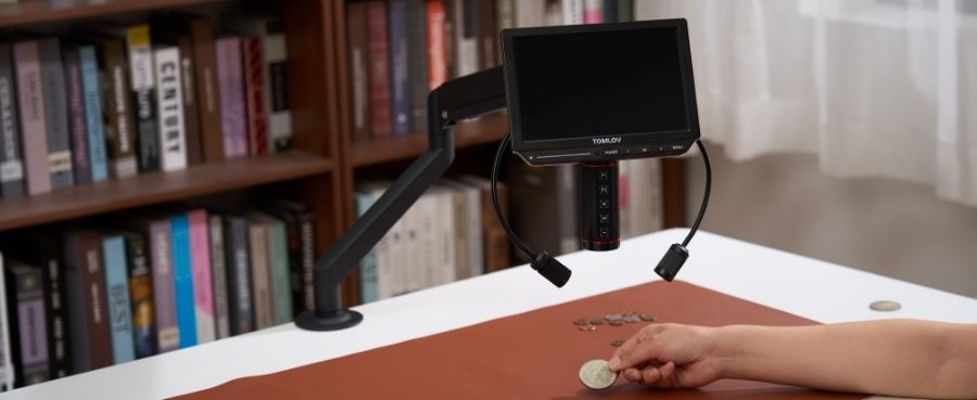
What Is the Proper Way to Carry a Microscope?
Learn the proper way to carry a microscope safely. Discover step-by-step techniques to support the base, protect delicate optics, and prevent damage
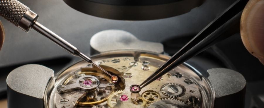
Microscope for Watch Repair: How to Use It for Tiny Watch Parts
Discover how a microscope simplifies watch repair. Get practical tips on inspecting gears, placing microscopic screws, and setting up working distance.
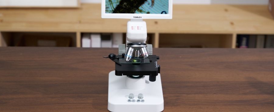
Numerical Aperture vs Magnification: How They Affect Microscope Resolution
Explore how numerical aperture and magnification control microscope resolution. Get simple tips to eliminate blur and improve image detail.
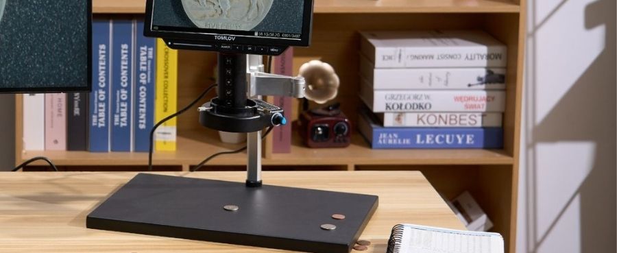
Polyester Under a Microscope: Fiber Shape, Texture, and Key Features
Explore what polyester looks like under a microscope. Discover its smooth, glass-like structure and learn how to compare it to cotton and wool.
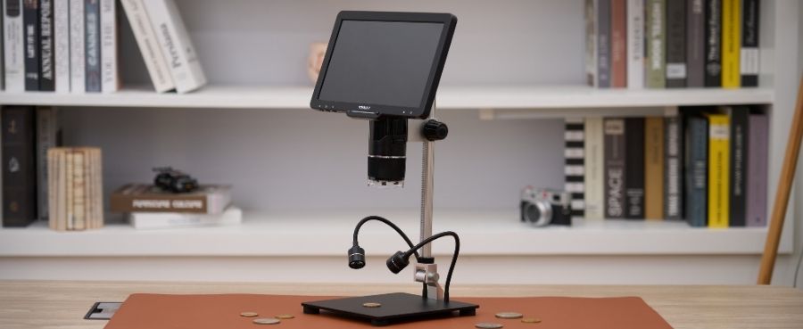
Function of a Stage on a Microscope: Simple Guide for Beginners
Understand the purpose of a microscope stage in this easy beginner's guide. Discover stage types, controls, and how to position slides for clear views.
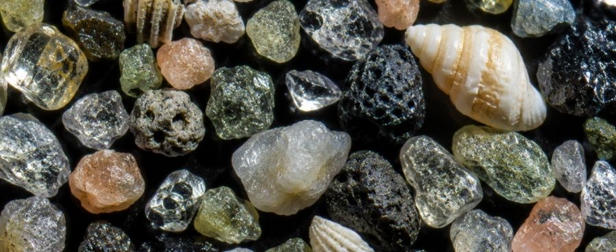
Sand Under a Microscope: What It Looks Like and How to Observe It
Step into the microscopic world of sand. Learn how grain shapes reveal geological history and get step-by-step tips for setting up your microscope.
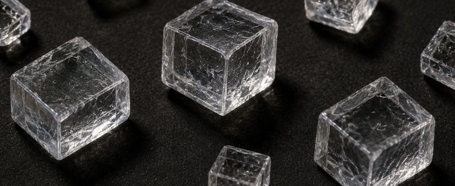
Salt Crystals Under a Microscope: What They Look Like
Uncover the cubic structure of salt under a microscope. We explain how to prepare samples, adjust lighting, and spot the differences between salt and sugar.
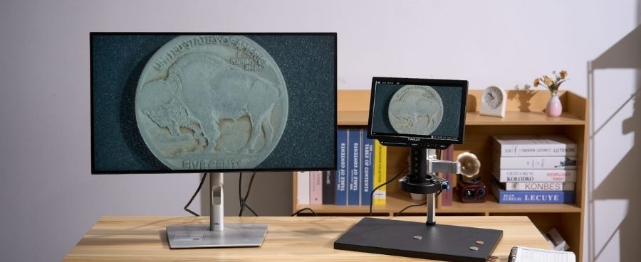
When Were Microscopes Invented? From Simple Lenses to Digital Microscopes
Discover when microscopes were invented, who shaped early microscopy, and how simple lenses evolved into today’s powerful digital microscopes.