
Table of Contents
Spirogyra is a green, thread-like freshwater alga that can show clear cell structure under a microscope. With a simple wet mount, you may see long filaments, repeated cell walls, spiral chloroplasts, and sometimes small pyrenoids. Start with a clean freshwater sample, trim a short filament, use one drop of water, and scan at low magnification before moving closer.
What Is Spirogyra and What Can You See Under a Microscope?
Green Filaments Made of Many Cells
Spirogyra often appears as soft green threads in ponds, lakes, ditches, or slow-moving streams. It is sometimes described as pond silk or blanket weed. Under a microscope, one green thread is actually a filament made of many connected cells. These cells form a repeated chain-like structure along the strand.
Spiral Chloroplasts Inside the Cells
The most recognizable feature of Spirogyra is the spiral chloroplast. These usually appear as bright green, ribbon-like bands winding through each cell. If the sample is thin and the focus is sharp, the spiral pattern can be easy to notice. This feature is one of the best beginner clues for recognizing Spirogyra.
Cell Walls, Cell Shape, and Visible Internal Detail
Spirogyra cells may look rectangular or cylindrical, depending on how the strand lies on the slide. Clear cell walls separate one cell from the next. At higher magnification, you may also see cross-walls, or septa, between neighboring cells. In a very clear sample, small pyrenoids may appear along the spiral chloroplasts.

What Should You Prepare Before Observing Spirogyra?
Look for Green Filamentous Algae in Freshwater
Look for soft green threads floating near the surface or attached to plants, rocks, or sticks. A pond, lake edge, shallow ditch, or slow-moving stream can be a good place to collect a sample. Not every green thread is Spirogyra, so the microscope observation is what confirms the visual clues.
Choose Clean, Fresh, and Safe Samples
Choose fresh samples from clean and safe freshwater sources. Avoid water that smells bad, looks polluted, or may contain chemical runoff. Wear gloves if needed and wash your hands after handling pond material. Collect only a small amount. A clean, thin strand is much easier to observe than a large tangled clump.
Prepare a Microscope With Bottom Light
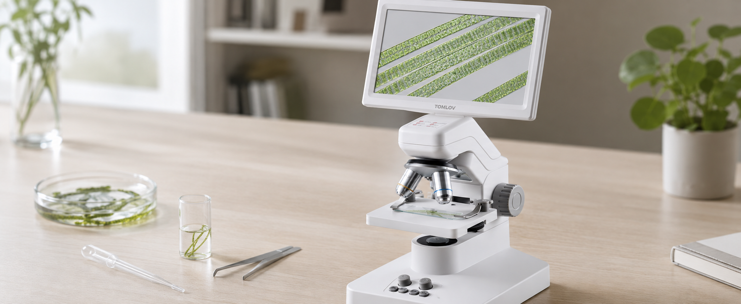
Spirogyra is usually viewed as a wet mount, so bottom light is useful because it passes through the slide, water, cover slip, and algae filament. For beginners, a digital microscope for specimen observation can make the process easier because you can view the sample on a screen while adjusting focus and lighting.
Slides, Cover Slips, Droppers, Tweezers, and Labels
Prepare clean slides, cover slips, a dropper, tweezers, paper towels, and labels. A dissecting needle or small scissors can also help if the filament is long or tangled. Label the sample with the collection place and date, especially if you plan to compare more than one freshwater sample.
How Do You Prepare and View a Spirogyra Wet Mount?
Place a Small Filament on the Slide
Use tweezers, a dropper, or a dissecting needle to pick up a small green thread. Spirogyra filaments can be several centimeters long, so trim or separate a short section before placing it on the slide. A short, untangled strand is easier to focus on than a long clump under the cover slip.
Add One Drop of Water
Add one small drop of water to keep the filament wet. Pond water from the original sample is usually best for a short observation. Water is enough for most beginner slides. A little glycerine can slow drying, but it is not required. Avoid too much liquid, because the filament may drift while you focus.
Lower the Cover Slip Gently
Hold the cover slip at about a 45-degree angle and lower it slowly over the drop. This helps reduce large trapped air bubbles. Do not press down on the cover slip. If the strand is too thick or tangled, make a thinner slide instead of forcing the sample flat.
Start at Low Magnification to Find the Filaments
Start at low power, around 40× total magnification if your microscope uses standard settings. Scan slowly until you find a clean, untangled strand. At this stage, Spirogyra may look like long green tubes joined end to end. Low magnification helps you separate real filaments from bubbles, debris, and other algae.
Increase Magnification to See Spiral Chloroplasts
After finding a clear filament, move to higher magnification, usually around 100× to 400×. Refocus slowly after each change. Look for cell walls first, then search inside the cells for spiral green chloroplasts. For students or beginners comparing several samples, the TOMLOV DM301 Pro digital microscope is useful here mainly because you can check the image on a screen and save photos while adjusting focus.
Save Clear Images Before the Slide Dries
Wet mounts can change quickly as water moves or dries. Save images while the filament is still flat, clear, and well lit. Capture one wider image showing the full filament chain, then one closer image showing the spiral chloroplasts and cell walls. A 4K digital microscope can be useful when you want sharper visual records for later comparison.

What Details Should Beginners Look For?
Long Chains of Rectangular or Cylindrical Cells
A good Spirogyra sample should show a long filament made of repeated cells. Follow one strand along its length and check whether the cell pattern continues. If the structure is random, broken, or not divided into cells, it may be debris or another type of algae.
Spiral Green Bands Inside Each Cell
The spiral green bands are the strongest beginner-level clue. These bands are chloroplasts, which help Spirogyra perform photosynthesis. If the spiral pattern is hard to see, adjust focus, reduce glare, or move to a thinner part of the strand before assuming the sample is not Spirogyra.
Clear Cell Walls Between Neighboring Cells
Look for thin lines between neighboring cells. These cross-walls, or septa, show where one cell ends and the next begins. Clear septa help you see that Spirogyra is built from many connected cells, not one continuous green tube.
Air Bubbles, Debris, and Other Microorganisms Nearby
Pond samples often contain bubbles, dirt, plant fragments, and small microorganisms. Air bubbles usually look round with dark edges. Tiny organisms may swim near the filament. These nearby details are normal, but Spirogyra itself should appear as a green filament made of connected cells.
What Mistakes Should Beginners Avoid?
Do Not Use Too Much Water on the Slide
Too much water makes the cover slip unstable and allows the filament to drift. It also makes focusing harder. Use one small drop first. If the slide begins to dry, add a tiny amount of water at the cover slip edge instead of flooding the slide.
Do Not Crush the Filaments Under the Cover Slip
Pressing the cover slip can flatten, break, or distort the filaments. This may hide the cell walls and spiral chloroplasts. Lower the cover slip gently and prepare a thinner slide if the sample looks too crowded.
Do Not Assume Every Green Thread Is Spirogyra
Many freshwater algae form green threads. Spirogyra is usually recognized by long filaments, repeated cells, clear cell walls, and spiral chloroplasts. If you see green threads but no spiral bands, the sample may be another kind of filamentous algae.
Do Not Overinterpret Species From One Slide
A beginner slide can help you recognize Spirogyra-like features, but it should not be used for exact species identification. Algae identification may require better samples, comparison references, and more expertise. Treat your slide as a visual observation, not a final classification.
Conclusion
Viewing Spirogyra under a microscope is a simple way to explore freshwater algae and basic cell structure. Collect a clean green filament, trim it to a short length, prepare a thin wet mount, and start at low magnification before moving closer. Look for long cell chains, clear septa, spiral chloroplasts, and sometimes pyrenoids. Keep the slide thin, avoid crushing the sample, and save images before the wet mount dries.
FAQs
How Does Spirogyra Reproduce Under a Microscope?
Spirogyra can reproduce by fragmentation or by sexual reproduction called conjugation. Under a beginner microscope, you may sometimes see broken filaments that could continue growing as separate pieces. Conjugation is harder to catch because it appears only under certain conditions and may show neighboring filaments connected by tube-like bridges.
How to Identify Spirogyra?
Spirogyra is usually recognized by long green filaments, chain-like cells, clear cell walls, and spiral chloroplasts inside the cells. The spiral green bands are the most distinctive beginner clue. However, not every green thread in pond water is Spirogyra, so use these features as visual clues rather than final species identification.
Can Spirogyra Move Under a Microscope?
Spirogyra does not swim like many small microorganisms. Under the microscope, the filaments may drift, bend, or shift because of water movement on the slide. If you see fast-moving organisms nearby, they are usually other pond-water microorganisms rather than Spirogyra itself.
Can You Use Tap Water for a Spirogyra Wet Mount?
Tap water can work for a short beginner observation, but pond water from the original sample is usually better. It keeps the algae in a more familiar environment during viewing. Use only a small drop on the slide. Too much water can make the cover slip float and push the filaments out of focus.

Leave a comment
All comments are moderated before being published.
This site is protected by hCaptcha and the hCaptcha Privacy Policy and Terms of Service apply.