Table of Contents
If you've ever wondered just how far a school microscope can actually zoom in, here's the short answer: most classroom compound light microscopes top out at 1,000x magnification. That's achieved by pairing a standard 10x eyepiece with a 100x oil-immersion objective lens. Some entry-level educational microscopes stop at 400x, while some advanced models advertise magnifications up to 2,000x. However, magnifications beyond about 1,000x often provide little additional detail due to the optical limits of visible light.
What Is a Classroom Compound Microscope?
A classroom compound microscope is the standard upright instrument you'll find in almost every middle school, high school, and intro-level college biology lab. It uses two sets of lenses — an eyepiece (ocular) and a rotating turret of objective lenses — to magnify thin, transparent specimens lit from below. "Compound" simply means the magnification happens in two stages instead of one, which is what lets these microscopes reach much higher powers than a single-lens magnifier ever could.
How does magnification actually work?
Magnification is just a measure of how much larger an object appears compared to its real size. In a compound microscope, light passes through the specimen, then through the objective lens (which produces an enlarged image), and finally through the eyepiece (which magnifies that image again before it reaches your eye). The total magnification is the product of those two stages — and that's why even modest individual lenses can combine to produce dramatic results. For students who want to skip the eyepiece entirely and view specimens on a built-in screen, a digital microscope follows the same two-stage principle but projects the magnified image directly onto a display.

Which parts determine the magnification?
The eyepiece lens
The eyepiece, or ocular, is the lens you look through at the top of the microscope. On nearly every classroom model, it's fixed at 10x magnification. Some advanced microscopes offer 15x or 20x eyepieces, but 10x is the universal standard in schools.
The objective lenses
The objectives sit on the rotating nosepiece just above the specimen. Classroom compound microscopes typically come with three or four of them: a 4x scanning objective for getting your bearings, a 10x low-power objective for general viewing, a 40x high-power objective for detailed cell work, and on more advanced models, a 100x oil-immersion objective for the highest magnification. Each one has its own focal length and resolving power, and they're color-coded by international convention so students can swap between them without confusion.
How the eyepiece and objective combine
Total magnification is just eyepiece × objective. With a 10x eyepiece, the math works out simply: 10x paired with the 4x objective gives 40x for scanning, 10x with the 10x objective gives 100x for low-power viewing, 10x with the 40x objective gives 400x for high-power work, and 10x with the 100x objective gives 1,000x using oil immersion. That last combination is the ceiling for most classroom work, and getting a usable image at 1,000x requires a drop of immersion oil between the lens and the slide to keep the light from scattering.
Magnification vs. Resolution: What's the Difference?
Magnification Makes the Image Larger
Magnification is about apparent size. A 1,000x magnification means the image appears one thousand times larger than the specimen in linear dimensions.That's it — no promise about clarity, just scale.
Resolution Reveals Fine Details
Resolution is the ability to distinguish two points that sit very close together. It's what determines whether a magnified image looks crisp or blurry. A high-magnification image with poor resolution is just a big blur, while a moderate-magnification image with excellent resolution can reveal far more useful detail. That's why, when picking a classroom microscope, what matters isn't the biggest number on the box — it's the quality of the optics behind it. A well-built 3-Lens Microscope that delivers sharp, clean images at 40x, 100x, and 400x will teach students far more than a cheap unit claiming 2,000x with a blurry view at every level.

What are the practical limits of classroom microscope magnification?
Empty magnification beyond 1000x
Past about 1,000x on a standard classroom light microscope, you begin to encounter what microscopists call "empty magnification."The image keeps getting bigger, but no new detail appears — it just turns into a softer, blurrier version of the same picture.
Resolution limited by the wavelength of light
This isn't a flaw in the lenses; it's physics. Visible light has a wavelength of roughly 400–700 nanometers, and you can't resolve details smaller than about half that wavelength no matter how good your optics are. This is why electron microscopes exist — they use electron beams with much shorter wavelengths to break past the limit.
Shrinking depth of field and working distance
At higher magnifications, the depth of field collapses — only an incredibly thin slice of the specimen stays in focus at once. The working distance (the gap between the lens and the slide) also shrinks dramatically, sometimes to less than a millimeter at 100x objective.
The need for stronger illumination
The more you magnify, the more light you need. At 1,000x, classroom microscopes rely on a bright built-in illuminator and a fully open condenser just to give you a viewable image. Insufficient illumination at high magnification can result in a dim image and loss of visible detail.
What can students actually see at each magnification level?
40x for getting the big picture
At 40x, you're seeing the overall shape and layout of a specimen. This is the magnification for locating things on the slide, scanning tissue sections, or examining larger structures like leaf cross-sections and small invertebrates.
100x for cells and tissues
At 100x, individual plant and animal cells become clearly visible. Onion skin cells, cheek cells, and tissue arrangements are perfect at this power.
400x for fine cell details
At 400x, you can see inside cells — nuclei, chloroplasts, vacuoles, and the movement of organelles in living specimens. Most of the "wow" moments in a biology class happen at this magnification.
1000x for bacteria and the smallest structures
At 1,000x with oil immersion, individual bacteria become resolvable. You can see their shapes (rods, spheres, spirals) and arrangements, which is essential for any microbiology unit. This is the practical ceiling for classroom light microscopy.
How can students get the clearest image at max magnification?
Start low and work your way up
Always begin at 40x to locate your specimen, then center it before switching to higher objectives. Trying to find something at 1,000x is like searching for a contact lens with a telescope.
Adjust lighting and focus carefully
Use the coarse focus only on low power. At 400x and above, switch to the fine focus knob — even a tiny adjustment shifts the entire view. Open the condenser and turn up the illuminator as you increase magnification.
Choose a microscope built for clarity
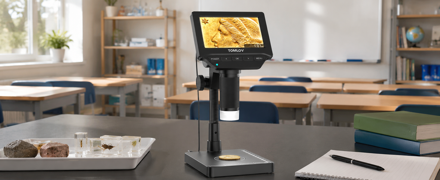
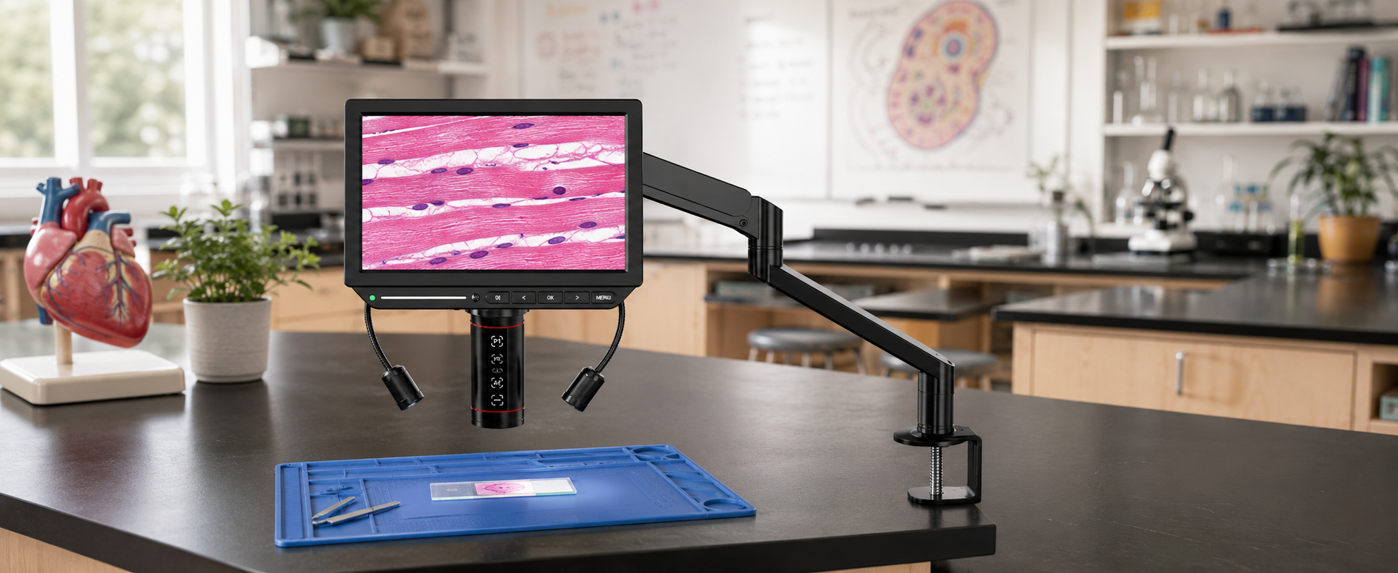
Technique only gets you so far if the optics aren't up to the job. A unit with quality glass, stable mechanics, and reliable lighting will produce noticeably better images at every magnification level — and increasingly, classrooms are pairing or replacing traditional compound microscopes with digital versions that work on the same two-lens principle but project the image onto a built-in screen instead of an eyepiece. This makes it far easier for students to share what they're seeing, record findings, and avoid the eye strain of long sessions at the eyepiece. The Tomlov digital microscope lineup is built around this approach, offering high-resolution imaging, stable LED illumination, and a magnification range that holds up clearly at the upper end where most classroom microscopes start to struggle.
Conclusion
The maximum magnification of a classroom compound light microscope is 1,000x — a number set by the combination of a 10x eyepiece and a 100x oil-immersion objective, and capped by the wavelength of visible light itself. Going higher with a standard light microscope just produces empty magnification: bigger, blurrier, no more useful. What matters far more than chasing big numbers is matching the right magnification to what you're trying to see, keeping your technique sharp, and using a microscope whose optics and lighting actually hold up at the upper end. Get those three things right, and a basic classroom microscope can take students from a leaf cross-section all the way down to a single bacterium.
FAQs
What is the maximum magnification of most classroom compound microscopes?
Most classroom compound microscopes max out at 1,000x, using a 10x eyepiece with a 100x oil-immersion objective lens.
What are 10x, 40x, and 100x objective lenses used for?
A 10x objective is commonly used for observing larger structures and tissues, a 40x objective is ideal for examining cellular details, and a 100x oil-immersion objective is used for viewing bacteria and other very small specimens at maximum magnification.
What is the maximum magnification of a microscope?
Light microscopes peak around 1,000–2,000x, while electron microscopes can reach over 2,000,000x using electron beams instead of light.
Do most compound microscopes have a 4x, 10x, and 40x objective lens?
Yes — 4x, 10x, and 40x is the standard objective set on nearly every classroom compound microscope, with 100x optional.
Why does my microscope image get darker at higher magnification?
Higher magnification illuminates a smaller area, so less light reaches your eye. Open the condenser and brighten the illuminator to compensate.
Hinterlasse einen Kommentar
Alle Kommentare werden vor der Veröffentlichung geprüft.
Diese Website ist durch hCaptcha geschützt und es gelten die allgemeinen Geschäftsbedingungen und Datenschutzbestimmungen von hCaptcha.