
Table of Contents
Blurry microscope images often stem from a limited depth of field. This article explains the core factors dictating image sharpness, including magnification and numerical aperture. You will discover practical ways to measure, adjust, and increase your focal range for clear observations.
What Is Depth of Field
Depth of field refers to the vertical distance or thickness of your specimen that remains perfectly in focus at one time. Imagine a transparent slice of space floating horizontally beneath your lens. Anything inside this specific slice appears crystal clear, while everything above or below it blurs into the background. A deep range lets you see thick objects clearly from top to bottom. A shallow range means only a paper-thin layer is visible at once.
Depth of Field Compared With Related Microscope Terms
Microscopy has a very specific vocabulary. Understanding the differences between these optical terms prevents confusion when you are trying to adjust your equipment.
Depth of Field and Depth of Focus
People often use these terms interchangeably, but they refer to completely different physical areas. Depth of field happens down at the specimen stage, representing the sharp area within your sample. Depth of focus happens up inside the camera sensor or eyepiece, representing the exact distance the image plane can move while still appearing sharp to the user.
Depth of Field and Field of View
While both terms describe what you see, they measure space in different directions. Field of view is the horizontal circle of light visible through the eyepiece, dictating image width. Depth of field measures the vertical thickness of the sharp area. Simply put, field of view is the overall width, and depth of field is the overall height of your focal area.
Depth of Field and Working Distance
Working distance is the physical gap between the bottom of your objective lens and the top of your glass slide. It tells you how much physical room you have to maneuver tools or manipulate the sample. Depth of field is strictly an optical measurement of image sharpness. However, lenses with very short working distances typically also produce a very shallow focal range.
What Determines Microscope Depth of Field?
Several physical and optical factors work together to dictate how much of your specimen stays sharp. Knowing these factors helps you control the final image.
Magnification
Magnification plays the largest role in determining how much of your sample stays sharp. As you zoom in on tiny details, the slice of focus naturally shrinks. A basic four times magnification lens provides a deep focal area for bulky items. Moving to a one hundred times magnification lens shrinks that sharp area to a tiny fraction of a millimeter.

Numerical Aperture
Numerical aperture measures how effectively your lens gathers light and resolves fine detail. High numerical aperture lenses capture incredible resolution at the direct expense of focal depth. The higher the numerical aperture printed on your objective lens, the thinner your slice of focus will become.
Aperture Diaphragm
The aperture diaphragm is the adjustable opening under your viewing stage that controls the angle of light hitting the sample. By closing this opening slightly, you force the light rays into a narrower cone. This narrow cone effectively increases your vertical focal range, though closing it too much can introduce visual artifacts or make the image entirely too dark.
Specimen Thickness
The physical nature of your sample impacts your viewing experience just as much as the lens. A single layer of blood cells smeared flat across a slide requires very little focal depth to appear sharp. Conversely, examining a bulky object like a mineral crystal requires a massive focal range to keep high peaks and deep valleys in focus simultaneously.
Specimen Position
How you position the specimen on the stage changes how the focal range interacts with it. If a flat object is slightly tilted, one edge might sit completely outside your sharp focal slice while the other remains clear. Ensuring your sample rests perfectly flat maximizes your available focal depth across the entire viewing area.
How to Estimate or Measure Microscope Depth of Field
You do not have to guess how much focal depth you have to work with. There are reliable ways to determine the exact thickness of your sharp viewing area.
Using Objective Specifications
The easiest way to estimate your focal range is to read the numbers printed directly on the barrel of your objective lens. By checking the magnification and numerical aperture, you can consult standard optical charts provided by the manufacturer to find the exact focal thickness designed for that specific lens.
Using a Depth-of-Field Formula
For absolute precision, you can calculate the exact thickness using a mathematical formula. The total depth involves adding the wave optical depth and the geometric depth together. This requires knowing the exact wavelength of your light source, the refractive index of the viewing medium, and the numerical aperture of your equipment.
Measuring Depth of Field Experimentally
You can measure your focal range manually using a specialized calibration slide featuring microscopic angled ridges. By bringing a specific section of the angled ridge into focus and reading the corresponding micrometers marked on the slide, you can physically test how much vertical space remains sharp at one time.
How to Increase Microscope Depth of Field
When your specimen is too thick for your current focal range, you can use a few practical techniques to squeeze more depth out of your optics.
Start With Lower Magnification
The most reliable way to increase your focal thickness is to simply use a lower power lens. Always begin your observations at the lowest possible magnification. This gives you a deep, forgiving focal area that makes it incredibly easy to find your subject before zooming in closer.
Adjust the Aperture Gradually
You can gain a noticeable boost in focal thickness by manipulating the condenser under your stage. Slowly close the aperture diaphragm while looking through the eyepiece. The image will become slightly darker but noticeably sharper from top to bottom. Just be careful not to close it entirely to avoid ruining the resolution.
Level the Specimen
If you cannot increase the optical focal range, you must adapt your specimen to fit within the existing slice of focus. Press your coverslip down gently to flatten the biological sample. Making the object as physically flat as possible ensures the entire surface area remains within your limited sharp zone.
Improve the Lighting
Bright and even illumination allows you to close the aperture diaphragm further without making the image too dark to see. Upgrade to a powerful LED light source or adjust your external lamps. Better lighting gives you the flexibility to tweak your optical settings for maximum focal thickness.
Prepare a Thinner Sample
Sometimes the absolute best solution is to modify the subject itself. Instead of trying to force thick plant tissue into focus, use a sharp razor blade to slice a much thinner cross-section. A thin sample eliminates the struggle of deep focal ranges and allows light to pass through perfectly.
Choosing the Right Microscope Setup
Different viewing subjects require completely different hardware setups. Matching your equipment to your sample solves most focal problems automatically.
Compound Microscopes for Thin Samples
Compound models are explicitly designed to view incredibly thin, transparent specimens mounted on glass slides. Because they use high-magnification lenses with high numerical apertures, their focal range is exceptionally shallow. They are the perfect tool for viewing single cellular organisms where deep focus is neither needed nor possible.
Stereo Microscopes for Three-Dimensional Objects
Stereo models operate at much lower magnifications, typically between ten and forty times power. This lower power provides a massive focal range, making them ideal for inspecting bulky objects like circuit boards, gemstones, and whole insects. They allow you to see the entire surface texture simultaneously.
Digital Microscopes for Image Stacking

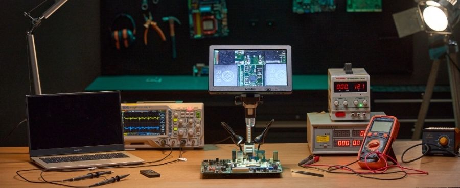
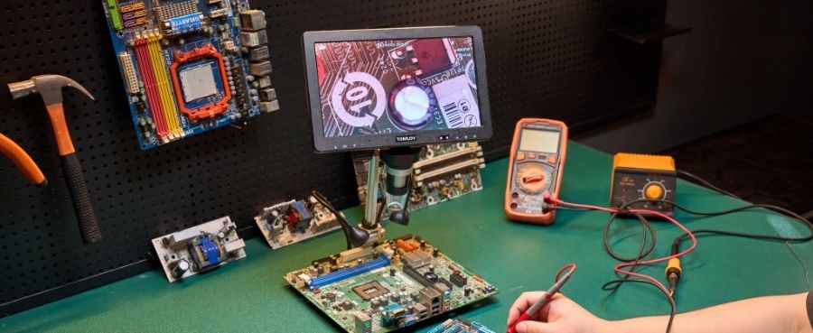
Modern digital microscopes solve focal limitations through advanced software. A system like Tomlov DM9 Max 10.1 HD IPS HDMI Digital Microscope allows you to capture crystal-clear video and images. Advanced digital software can take multiple photos at different focal heights and merge them together, creating a single photograph where a thick object is entirely sharp from front to back.
Conclusions
Mastering your focal range fundamentally changes how you interact with microscopic specimens. By understanding the strict relationship between magnification, numerical aperture, and your hardware settings, you can permanently stop struggling with blurry edges. Whether you adjust your lighting, prepare thinner samples, or switch to a digital system equipped for focus stacking, you now have the exact knowledge required to capture incredibly sharp and professional results every single time you observe a sample.
Frequently Asked Questions
How do you calculate the FOV of a microscope?
You can calculate the field of view by dividing the field number printed on your eyepiece by the magnification of your objective lens. For example, if your eyepiece has a field number of twenty and you use a ten times objective, your field of view is exactly two millimeters wide.
How to find depth of field microscope?
You find the available focal thickness by identifying the numerical aperture on your lens and consulting standard optical reference tables. Alternatively, you can close your aperture diaphragm slightly to visually find the maximum vertical space your specific lens can keep sharp on a known specimen.
How to measure the field of view?
The most practical way to measure the field of view is to place a transparent metric ruler under the lens. Count the number of millimeter lines visible across the center of the illuminated circle to find the exact physical width of your viewing area.
Does the 10x or 40x have a shorter depth of field?
A forty times lens has a significantly shorter focal range than a ten times lens. Because the forty times lens provides higher magnification and features a larger numerical aperture, the slice of sharp focus becomes incredibly thin compared to the lower power option.
Why Depth of Field Decreases at High Magnification?
Focal thickness naturally decreases at high magnification because the lens must gather light from a much wider angle to resolve tiny details. This wider angle of light collection physically narrows the vertical band where the light rays converge perfectly into a sharp image.


Hinterlasse einen Kommentar
Alle Kommentare werden vor der Veröffentlichung geprüft.
Diese Website ist durch hCaptcha geschützt und es gelten die allgemeinen Geschäftsbedingungen und Datenschutzbestimmungen von hCaptcha.